Japanese
English
- 有料閲覧
- Abstract 文献概要
- 1ページ目 Look Inside
- サイト内被引用 Cited by
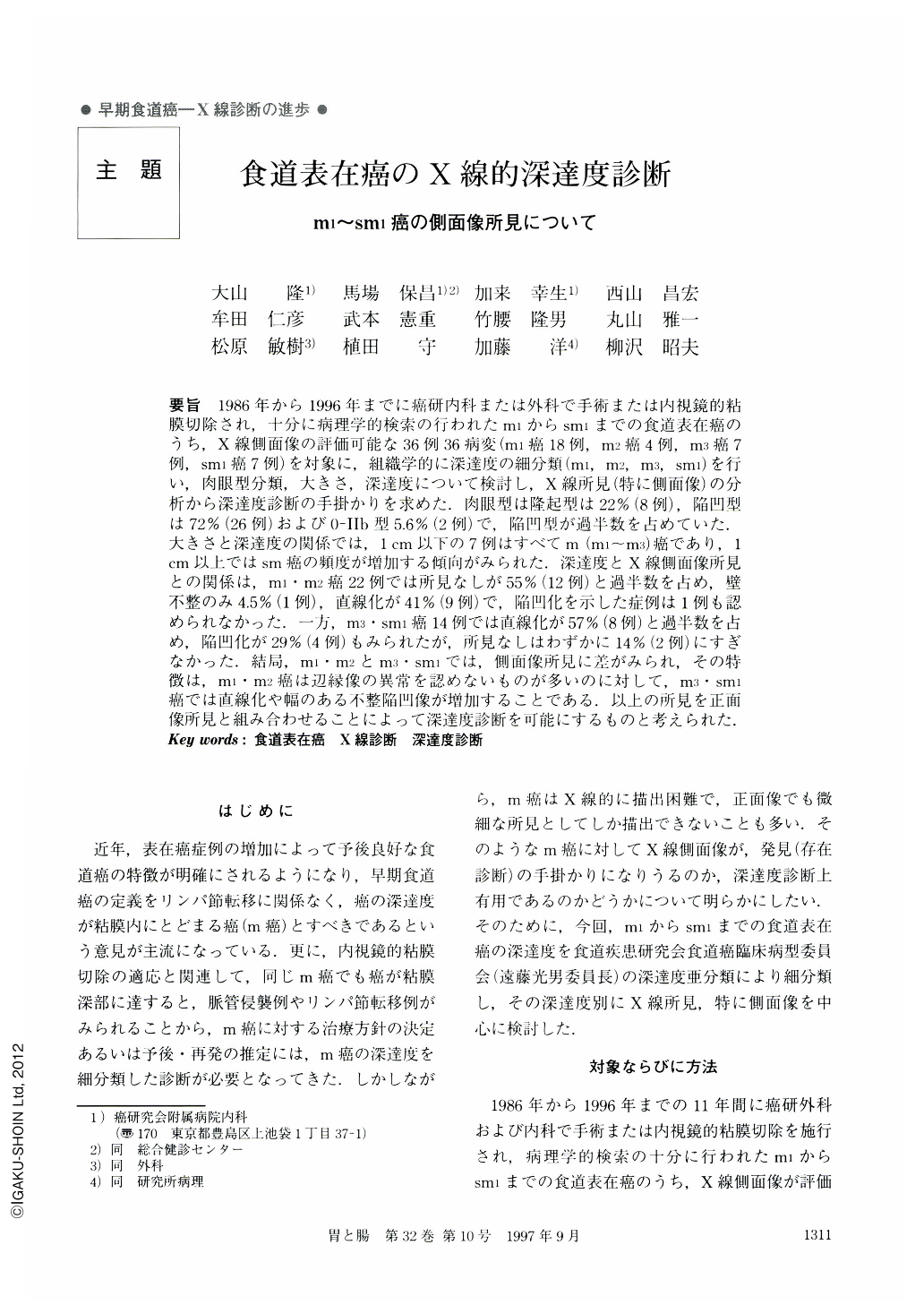
要旨 1986年から1996年までに癌研内科または外科で手術または内視鏡的粘膜切除され,十分に病理学的検索の行われたm1からsm1までの食道表在癌のうち,X線側面像の評価可能な36例36病変(m1癌18例,m2癌4例,m3癌7例,sm1癌7例)を対象に,組織学的に深達度の細分類(m1,m2,m3,sm1)を行い,肉眼型分類,大きさ,深達度について検討し,X線所見(特に側面像)の分析から深達度診断の手掛かりを求めた.肉眼型は隆起型は22%(8例),陥凹型は72%(26例)および0-Ⅱb型5.6%(2例)で,陥凹型が過半数を占めていた.大きさと深達度の関係では,1cm以下の7例はすべてm(m1~m3)癌であり,1cm以上ではsm癌の頻度が増加する傾向がみられた.深達度とX線側面像所見との関係は,m1・m2癌22例では所見なしが55%(12例)と過半数を占め,壁不整のみ4.5%(1例),直線化が41%(9例)で,陥凹化を示した症例は1例も認められなかった.一方,m3・sm1癌14例では直線化が57%(8例)と過半数を占め,陥凹化が29%(4例)もみられたが,所見なしはわずかに14%(2例)にすぎなかった.結局,m1・m2とm3・sm1では,側面像所見に差がみられ,その特徴は,m1・m2癌は辺縁像の異常を認めないものが多いのに対して,m3・sm1癌では直線化や幅のある不整陥凹像が増加することである.以上の所見を正面像所見と組み合わせることによって深達度診断を可能にするものと考えられた.
During the period between 1986 and April 1996, there were 36 lesions from 36 cases of superficial esophageal cancer (18 cases of the m1 cancer, four cases of the m2 cancer, seven cases of the m3 cancer, and seven cases of the sm1 cancer) that were resected by operation or endoscopic mucosal resection and could be eveluated by the lateral view of the radiologic examination in the medical and surgical department of the Cancer Research Hospital. Those lesions were analyzed for the evaluation of usefulness of lateral view of the radiologic examination from the viewpoints of the macroscopic classification, size and depth of invasion. Macroscopically, the depressive type consisted more than half of the lesions (72%, 26 cases) compared to the elevated type (22%, eight cases) and the Ⅱb type (5.6%, two cases). Concerning the relationship between size of the lesion and depth of invasion, all seven cases of the lesion less than 1 cm in size were m (m1~ m3) cancer. The incidence of the sm1 cancer might be increased with the size of the lesion when it was 1 cm or larger. The relationships between the depth of lesion and radiologic lateral view were as follows: in the m1 and m2 cancer group (22 cases), no abnormal findings, 55 (12 cases); wall irregularity, 2.8% (one case); and wall straight change 41% (eight cases). In the m3 and sm1 cancer group (14 cases), wall straight change, 57% (eight cases); depression, 29% (four cases); and no abnormal findings, 14% (two cases). In summary, there were differences of lateral view findings between the group of m1 and m2 cancers and the group of m3 and sm1 cancers: more than half of the m1 and m2 cancer did not have abnormal findings on the lateral view, and m3 and sm1 cancer might have wall straight change and wide irregular depressive image. Combination of frontal and lateral views would help to diagnose the depth of invasion.

Copyright © 1997, Igaku-Shoin Ltd. All rights reserved.


