Japanese
English
- 有料閲覧
- Abstract 文献概要
- 1ページ目 Look Inside
- サイト内被引用 Cited by
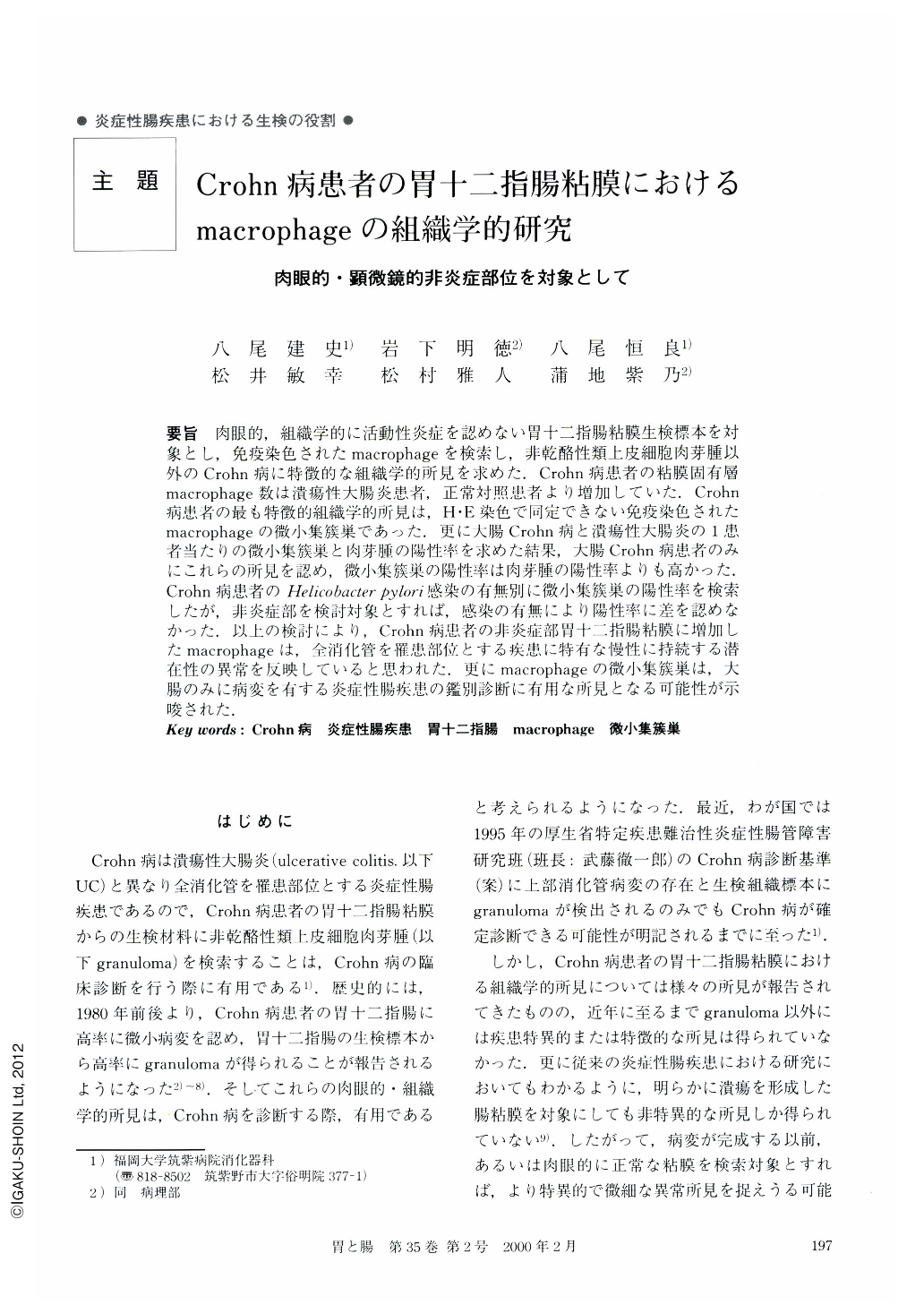
要旨 肉眼的,組織学的に活動性炎症を認めない胃十二指腸粘膜生検標本を対象とし,免疫染色されたmacrophageを検索し,非乾酪性類上皮細胞肉芽腫以外のCrohn病に特徴的な組織学的所見を求めた.Crohn病患者の粘膜固有層macrophage数は潰瘍性大腸炎患者,正常対照患者より増加していた.Crohn病患者の最も特徴的組織学的所見は,H・E染色で同定できない免疫染色されたmacrophageの微小集簇巣であった.更に大腸Crohn病と潰瘍性大腸炎の1患者当たりの微小集簇巣と肉芽腫の陽性率を求めた結果,大腸Crohn病患者のみにこれらの所見を認め,微小集籏巣の陽性率は肉芽腫の陽性率よりも高かった.Crohn病患者のHelicobacter pyori感染の有無別に微小集籏巣の陽性率を検索したが,非炎症部を検討対象とすれば,感染の有無により陽性率に差を認めなかった.以上の検討により,Crohn病患者の非炎症部胃十二指腸粘膜に増加したmacrophageは,全消化管を罹患部位とする疾患に特有な慢性に持続する潜在性の異常を反映していると思われた.更にmacrophageの微小集簇巣は,大腸のみに病変を有する炎症性腸疾患の鑑別診断に有用な所見となる可能性が示唆された.
We investigated the characteristic histological findings of immunostained macrophages in noninflamed gastroduodenal mucosa of patients with Crohn's disease, ulcerative colitis and healthy control. The number of immunostained macrophages in Crohn's disease patients was increased than those in ulcerative colitis and control patients. The most characteristic finding was a microaggregate of immunostained macrophages which had not been visualized by H・E staining alone. No microaggregate was detected in either ulcerative colitis or control patients. The incidence of microaggregate of macrophages in Crohn's disease patients has no relationship to Helicobacter pylori infection. In conclusion, the histological finding of focally accumulated macrophages with microaggregate in noninflamed gastroduodenal mucosa might be a persistent latent abnormality, reflecting the entire involvement of the alimentary tract in Crohn's disease. In addition, detecting a microaggregate of immunostained macrophages in noninflamed gastroduodenal mucosa might be a useful marker for differentiating Crohn's disease from ulcerative colitis.

Copyright © 2000, Igaku-Shoin Ltd. All rights reserved.


