Japanese
English
- 有料閲覧
- Abstract 文献概要
- 1ページ目 Look Inside
- 参考文献 Reference
- サイト内被引用 Cited by
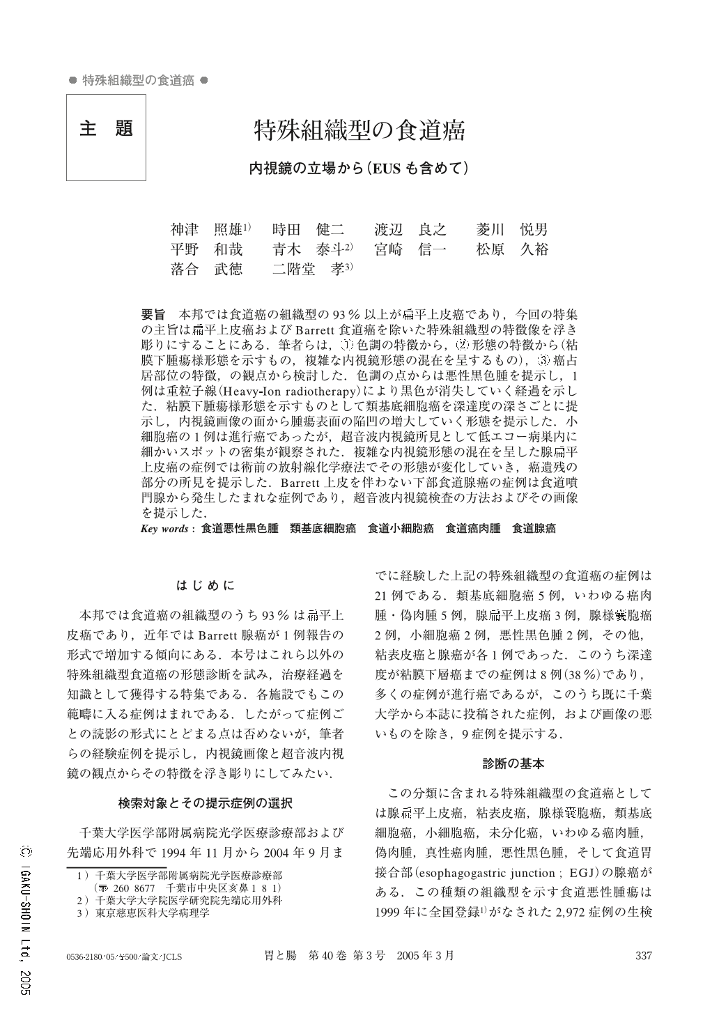
要旨 本邦では食道癌の組織型の93%以上が扁平上皮癌であり,今回の特集の主旨は扁平上皮癌およびBarrett食道癌を除いた特殊組織型の特徴像を浮き彫りにすることにある.筆者らは,①色調の特徴から,②形態の特徴から(粘膜下腫瘍様形態を示すもの,複雑な内視鏡形態の混在を呈するもの),③癌占居部位の特徴,の観点から検討した.色調の点からは悪性黒色腫を提示し,1例は重粒子線(Heavy-Ion radiotherapy)により黒色が消失していく経過を示した.粘膜下腫瘍様形態を示すものとして類基底細胞癌を深達度の深さごとに提示し,内視鏡画像の面から腫瘍表面の陥凹の増大していく形態を提示した.小細胞癌の1例は進行癌であったが,超音波内視鏡所見として低エコー病巣内に細かいスポットの密集が観察された.複雑な内視鏡形態の混在を呈した腺扁平上皮癌の症例では術前の放射線化学療法でその形態が変化していき,癌遺残の部分の所見を提示した.Barrett上皮を伴わない下部食道腺癌の症例は食道噴門腺から発生したまれな症例であり,超音波内視鏡検査の方法およびその画像を提示した.
In Japan, squamous cell carcinomas verified by histological analyses account for some 93 % of the cases of esophageal carcinoma, so, in the current project, we underscored the characteristic images of rare cases of esophageal carcinoma excluding squamous cell carcinoma and Barrett's esophageal carcinoma. We investigated rare cases of esophageal carcinoma, from the standpoint of their specific ; ( 1 ) color tone, ( 2 ) morphology of (such as submucosal tumor and coexistence of comflicated endoscopic features, and ( 3 ) location of the carcinoma. Regarding the color tone, we presented a case of malignant melanoma and a process during which the black color disappeared due to Heavy-Ion Radiotherapy. As a case looking like a submucosal tumor, we presented cases of basaloid carcinoma and its stages of invasion depth seen in endoscopic images showing the morphological development of the surface depression. As a case of small cell carcinoma, which was actually advanced cancer, EUS visualized dense small spots in the low echoic seat of the disease. As a case of adenosquamous carcinoma showing the co-existence of comflicated endoscopic features we presented findings of a residual part of a carcinoma whose morphology had been changed by preoperative Chemo-Radio-Therapy. As a case of adenocarcinoma without Barrett's epithelium detected in the lower esophagus, we presented a rare case which had generated from the esophagocardiac gland and we explained how to visualize the lesion by EUS and through images taken during EUS.

Copyright © 2005, Igaku-Shoin Ltd. All rights reserved.


