Japanese
English
- 有料閲覧
- Abstract 文献概要
- 1ページ目 Look Inside
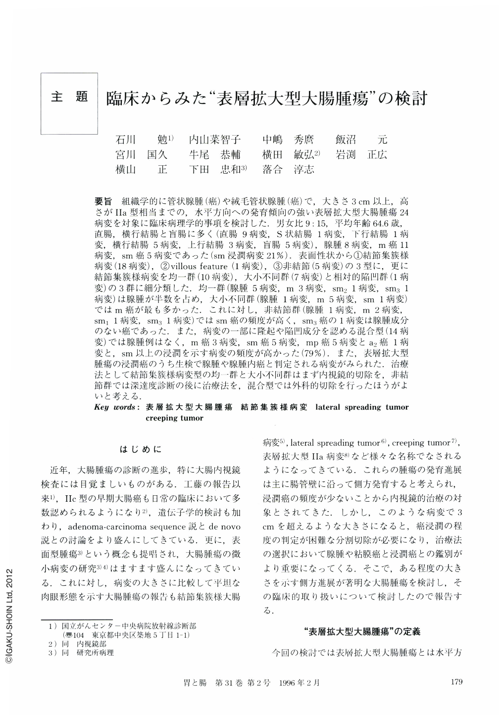
要旨 組織学的に管状腺腫(癌)や絨毛管状腺腫(癌)で,大きさ3cm以上,高さがⅡa型相当までの,水平方向への発育傾向の強い表層拡大型大腸腫瘍24病変を対象に臨床病理学的事項を検討した.男女比9:15,平均年齢64.6歳,直腸,横行結腸と盲腸に多く(直腸9病変,S状結腸1病変,下行結腸1病変,横行結腸5病変,上行結腸3病変,盲腸5病変),腺腫8病変,m癌11病変,sm癌5病変であった(sm浸潤病変21%).表面性状から①結節集簇様病変(18病変),②villous feature(1病変),③非結節(5病変)の3型に,更に結節集簇様病変を均一群(10病変),大小不同群(7病変)と相対的陥凹群(1病変)の3群に細分類した.均一群(腺腫5病変,m3病変,sm11病変,sm31病変)は腺腫が半数を占め,大小不同群(腺腫1病変,m5病変,sm1病変)ではm癌が最も多かった.これに対し,非結節群(腺腫1病変,m2病変,sm11病変,sm31病変)ではsm癌の頻度が高く,sm3癌の1病変は腺腫成分のない癌であった.また,病変の一部に隆起や陥凹成分を認める混合型(14病変)では腺腫例はなく,m癌3病変,sm癌5病変,mp癌5病変とa2癌1病変と,sm以上の浸潤を示す病変の頻度が高かった(79%).また,表層拡大型腫瘍の浸潤癌のうち生検で腺腫や腺腫内癌と判定される病変がみられた.治療法として結節集簇様病変型の均一群と大小不同群はまず内視鏡的切除を,非結節群では深達度診断の後に治療法を,混合型では外科的切除を行ったほうがよいと考える.
The purpose of this paper is to analyze the clinicopathological features in 24 cases in National Cancer Center Hospital of superficial spreading tumor (the height of the lesions is extremely low compared with the size of the entire lesion, larger than 2 cm in size) of the large intestine.
The average age of these cases was 64.6-year-old. Male to female ratio was nine to 15. Nine lesions were located in the rectum, five in the transverse colon and the cecum, three in the ascending colon and one in the sigmoid and descending colon, respectively. The pathological diagnosis were adenoma in eight lesions, mucosal cancer (m) in adenoma in 11 lesions, submucosal cancer (sm) in adenoma in four lesions, and sm without adenoma in one lesion.
The indications for endoscopic mucosal resection are Type Ⅰ and Ⅱ, but Type Ⅲ may be considered as invasive carcinoma. We have to diagnose the depth of invasion correctly and select an adequate management, which include a surgical operation, for the treatment of superficial spreading tumors.
Superficial spreading tumors were classified into three types for the variation of the surface structure, and 1) Nodular type (18 lesions): nodule-aggregating tumor. These were furthermore subclassified into three groups for the variation of the size and arrangement of the nodules. Group Ⅰ (10 lesions): The size of each nodule is uniform. Group Ⅱ (seven lesions): The size of each nodule is multiform, large or small. Group Ⅲ (one lesion): The center of the lesion is depressed but shows fine nodular change. 2) Villous tumor type (one lesion): The surface of tumor shows villous features. 3) Non-nodular type (five lesions); Non-nodular surface of tumor. The incidence of sm cancer in nodular type was 20% in group Ⅰ (adenoma: 5, m: 3, sm2: 1, sm3: 1), 14% in group Ⅱ (adenoma: 1, m: 5, sm: 1), and 40% in group Ⅲ (adenoma: 1, m: 2, sm1: 1, sm3: 1). Endoscopic mucosal resection is indicated for group Ⅰ and Ⅱ. Group Ⅲ was considered as naving a high incidence of invasive carcinoma and it is necessary to diagnose the depth of invasion correctly and select adequate management.
In superficial spreading tumor with distinct depressed or protruded areas (14 lesions), there were 11 lesions of invasive carcinoma (m: 3, sm: 5, mp: 5, a2: 1) and we considered it is necessary to select a surgical operation for the treatment of these tumors.

Copyright © 1996, Igaku-Shoin Ltd. All rights reserved.


