Japanese
English
- 有料閲覧
- Abstract 文献概要
- 1ページ目 Look Inside
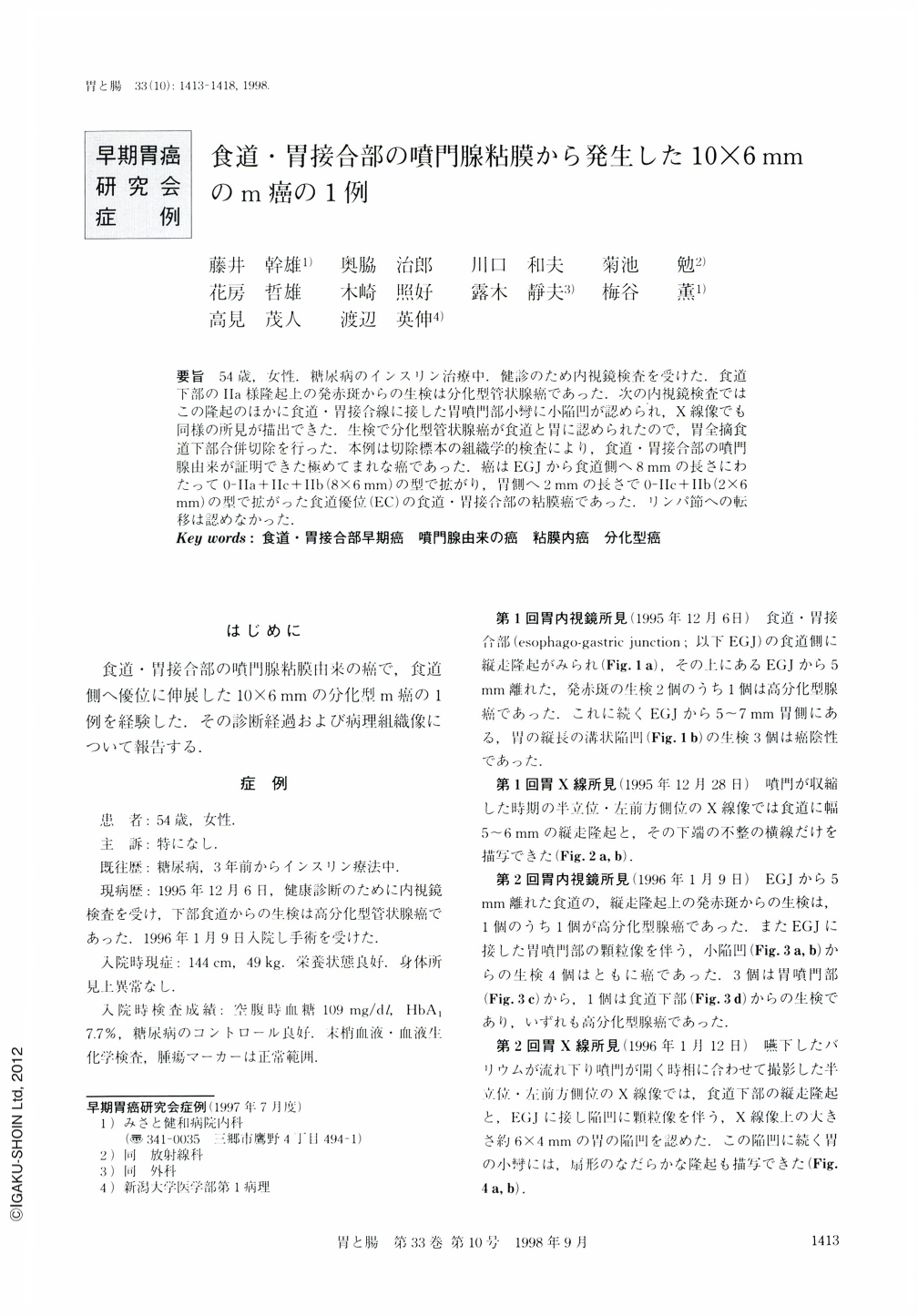
要旨 54歳,女性.糖尿病のインスリン治療中.健診のため内視鏡検査を受けた.食道下部のⅡa様隆起上の発赤斑からの生検は分化型管状腺癌であった.次の内視鏡検査ではこの隆起のほかに食道・胃接合線に接した胃噴門部小轡に小陥凹が認められ,X線像でも同様の所見が描出できた.生検で分化型管状腺癌が食道と胃に認められたので,胃全摘食道下部合併切除を行った.本例は切除標本の組織学的検査により,食道・胃接合部の噴門腺由来が証明できた極めてまれな癌であった.癌はEGJから食道側へ8mmの長さにわたって0-Ⅱa+Ⅱc+Ⅱb(8×6mm)の型で拡がり,胃側へ2mmの長さで0-Ⅱc+IIb(2×6mm)の型で拡がった食道優位(EC)の食道・胃接合部の粘膜癌であった.リンパ節への転移は認めなかった.
A 54-year-old woman had received insulin therapy for three years because of diabetes mellitus. X-ray examination of the esophagogastric junctuon (EGJ) in the left anterior lateral view, semistanding position revealed a fold-like mucosal elevated lesion of the esophagus and irregular shaped gastric mucosal depression with two tiny granules just below the EGJ. This was illustrated by swallowing contrast medium when the cardia had opened.
Endoscopic examination showed a fold-like elevation with a shallow reddish erosion on its surface in the lower end of the esophagus. Close-up endoscopic examination using the J-turn method showed a depression with irregular contour and small granules in this lesion. The biopsy specimen obtained from these two lesions in the EGJ region was positive for differentiated tubular adenocarcinoma.
Although this type of cancer development is very rare, histopathological examination of the resected specimen showed that differentiated adenocarcinoma had developed from the fundic glands located in the mucosa of EGJ region.
Cancer was also located in the EGJ region. It was composed of early mucosal cancer (0-IIc + IIb) measuring 2 × 6 mm in size in the lesser curvature of the stomach just below the EGJ, and early mucosal cancer (0-IIa + IIc + IIb) measuring 8 × 6 mm invading the esophageal mucosa. However, there was no metastasis to the lymph nodes.

Copyright © 1998, Igaku-Shoin Ltd. All rights reserved.


