Japanese
English
- 有料閲覧
- Abstract 文献概要
- 1ページ目 Look Inside
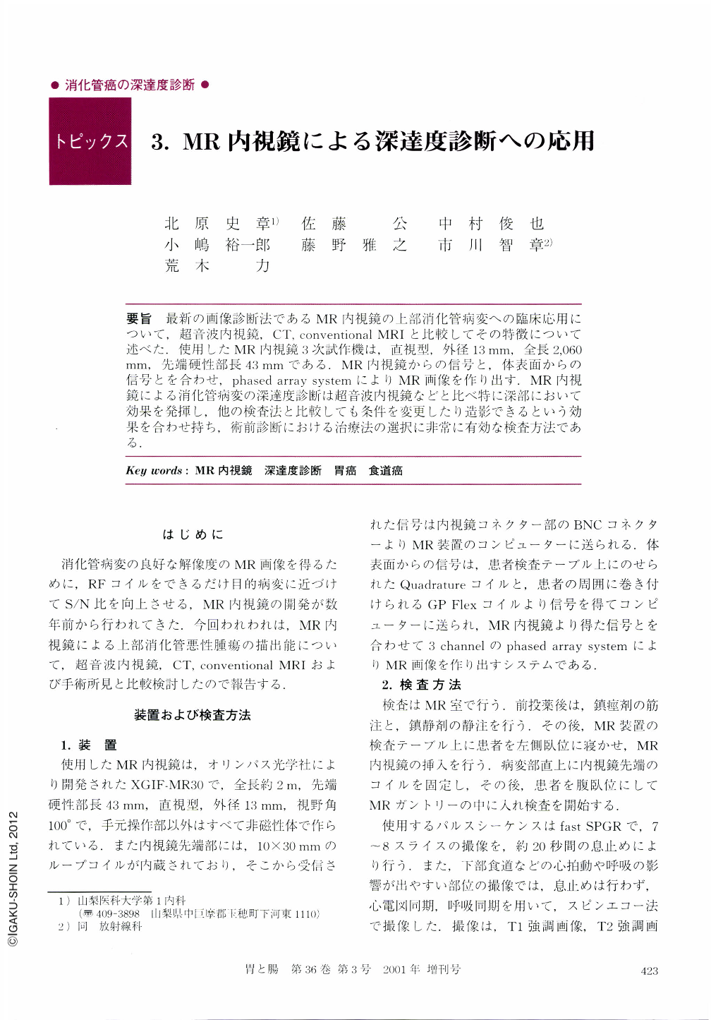
要旨 最新の画像診断法であるMR内視鏡の上部消化管病変への臨床応用について,超音波内視鏡,CT,conventional MRIと比較してその特徴について述べた.使用したMR内視鏡3次試作機は,直視型,外径13mm,全長2,060mm,先端硬性部長43mmである.MR内視鏡からの信号と,体表面からの信号とを合わせ,phased array systemによりMR画像を作り出す.MR内視鏡による消化管病変の深達度診断は超音波内視鏡などと比べ特に深部において効果を発揮し,他の検査法と比較しても条件を変更したり造影できるという効果を合わせ持ち,術前診断における治療法の選択に非常に有効な検査方法である.
Conventional magnetic resonance (MR) imaging of esophageal and gastric cancer is performed with body and surface coils. Body and surface coils often inadequately depict invasion into the esophageal adventitia or the gastric serosa. We performed local staging of esophageal and gastric cancer using an MR endoscope with a 3 cm-long receive-only coil embedded in its tip. MR endoscopy was performed with T1-weighted, fast-spin-echo T2-weighted, and frequency-selective fat-satu-rated T1-weighted gadolinium enhanced sequences. We are also investigating the optimal combination of a phased-array surface coil with the endoscopic coil to improve signal-to-noise ratio in our images. Esophageal cancer was imaged by a high-intensity display on Tl-weighted imaging and fast spin-echo. Gastric cancer was imaged by a low-intensity display on T1-weighted imaging and fast SPGR. Compared to endoscopic ultrasonography, we obtained clearer images of esophageal and gastric cancer in the deep layer by MR endoscopy. The advantages of endoscopic MRI are as follows: The three-dimensional tomography provided by this method enables us to select the area to be scanned, produces images that differ from those obtained with endoscopic ultrasonography or CT scanning, and allows us to evaluate the character of the tumor tissue by changing pulse sequence parameters, selecting adequate gradient-echo sequences, or using contrast media. We concluded that there are important potential advantages using this technique in lesion staging and patient treatment planning.

Copyright © 2001, Igaku-Shoin Ltd. All rights reserved.


