Japanese
English
- 有料閲覧
- Abstract 文献概要
- 1ページ目 Look Inside
- 参考文献 Reference
- サイト内被引用 Cited by
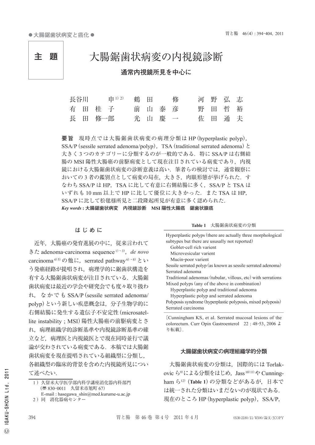
要旨 現時点では大腸鋸歯状病変の病理分類はHP(hyperplastic polyp),SSA/P(sessile serrated adenoma/polyp),TSA(traditional serrated adenoma)と大きく3つのカテゴリーに分類するのが一般的である.特にSSA/Pは右側結腸のMSI陽性大腸癌の前駆病変として現在注目されている病変であり,内視鏡における大腸鋸歯状病変の診断意義は高い.筆者らの検討では,通常観察においての3者の鑑別点として病変の局在,大きさ,肉眼形態が挙げられた.すなわちSSA/PはHP,TSAに比して有意に右側結腸に多く,SSA/PとTSAはいずれも10mm以上でHPに比して優位に大きかった.またTSAはHP,SSA/Pに比して松毬様所見と二段隆起所見が有意に多く認められた.
Today, the histopathology of colonic serrated lesions is generally classified grossly into three categories : HP(hyperplastic polyp), SSA/P(sessile serrated adenoma/polyp), and TSA(traditional serrated adenoma). As SSA/P has been recognized as antecedent of MSI-positive carcinoma of the right colon, endoscopic determination of colonic serrated lesions has a significant clinical role. Our study suggested that the three lesions can be differentiated by observation taking into account localization of the lesion, size, and macroscopic findings by conventional endoscopy. SSA/P was detected more frequently in the right colon than HP or TSA. Both SSA/P and TSA were also more than 10mm in diameter, which was significantly larger than HP. In addition, TSA was showed a significantly greater incidence of pine-cone like structure and double elevation than HP or SSA/P.
Copyright © 2011, Igaku-Shoin Ltd. All rights reserved.