Japanese
English
- 有料閲覧
- Abstract 文献概要
- 1ページ目 Look Inside
- 参考文献 Reference
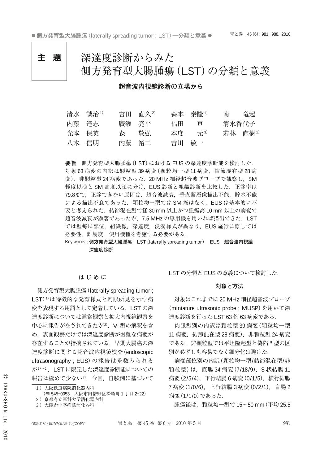
要旨 側方発育型大腸腫瘍(LST)におけるEUSの深達度診断能を検討した.対象63病変の内訳は顆粒型39病変(顆粒均一型11病変,結節混在型28病変),非顆粒型24病変であった.20MHz細径超音波プローブで観察し,SM軽度以浅とSM高度以深に分け,EUS診断と組織診断を比較した.正診率は79.8%で,正診できない原因は,超音波減衰,垂直断層像描出不能,貯水不能による描出不良であった.顆粒均一型ではSM癌はなく,EUSは基本的に不要と考えられた.結節混在型で径30mm以上かつ腫瘍高10mm以上の病変で超音波減衰が顕著であったが,7.5MHzの専用機を用いれば描出できた.LSTでは型毎に部位,組織像,深達度,浸潤様式が異なり,EUS施行に際しては必要性,難易度,使用機種を考慮する必要がある.
We retrospectively evaluated the efficacy of EUS for diagnosis of the infiltration depth of colorectal laterally-spreading tumors. The subjects were 63 lesions including 39 lesions of granular type(homogenous 11, nodular mixed 28)and 24 lesions of non-granular type. A 20MHz miniature ultrasonic probe(MUSP)was used for observation in all lesions, and a 7.5MHz ultrasonic endoscope was used for 23 lesions at the same time. The depth of invasion was divided into SM-slight>and SM-massive≦, and the results of EUS and histological diagnoses were compared. The infiltration depth was correctly diagnosed by MUSP in 79.8%. The cause of diagnostic failure in diagnosis was poor visualization. In granular type lesions, those with submucosal or deeper invasion were not included, and EUS diagnoses were correct except for one cecal lesion. For granular type lesions, EUS is considered unnecessary. The major reason for poor visualization was echo attenuation which was most remarkable in nodular mixed type of granular lesions larger than 30mm in diameter and 10mm in thickness. Such lesions, however, could be sufficiently visualized with the 7.5MHz echoendoscope. Other causes of poor visualization included inability to obtain perpendicular cross-sectional images of the lesions or to fill the lumen with deaerated water. These tendencies were often observed in the transverse or sigmoid colon. Since the histology, infiltration depth, mode of invasion and site of lesion differ according to the macroscopic type of lesions, the needs and difficulty in using EUS should be considered and an appropriate instrument should be selected.

Copyright © 2010, Igaku-Shoin Ltd. All rights reserved.


