Japanese
English
- 有料閲覧
- Abstract 文献概要
- 1ページ目 Look Inside
- 参考文献 Reference
- サイト内被引用 Cited by
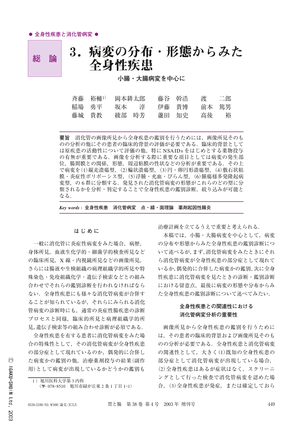
消化管の画像所見から全身疾患の鑑別を行うためには,画像所見そのものの分析の他にその患者の臨床的背景の評価が必要である.臨床的背景としては原疾患の活動性について評価の他,特にNSAIDsをはじめとする薬物投与の有無が重要である.画像を分析する際に重要な項目としては病変の発生部位,腸間膜との関係,形態,周辺粘膜の性状などの分析が重要である.その上で病変を(1)縦走潰瘍型,(2)輪状潰瘍型,(3)円・卵円形潰瘍型,(4)敷石状粘膜・炎症性ポリポーシス型,(5)浮腫・充血・びらん型,(6)腫瘍様多発隆起病変型,の6群に分類する.発見された消化管病変の形態がこれらのどの型に分類されるかを分析・判定することで全身性疾患の鑑別診断,絞り込みが可能となる.
In order to make a diagnosis of systemic disease from an analysis of shape and distribution of GI lesions, it is important to analyze the GI lesions as well as the patient's clinical background. A patient's clinical background, the activity of the original systemic disease and whether the patient uses drugs such as non-steroidal anti-inflammatory drugs are important sources of information. For a diagnosis of GI lesions obtained by radiologic or endoscopic examinations, it is important to analyze the location, shape and the properties of the surrounding mucosa of the lesions. GI lesions associated with systemic disease can be divided into the following six patterns proposed by Watanabe, et al. : i) longitudinal ulcer type ; ii) circular ulcer type ; iii) round or oval ulcer type ; iv) cobblestone or inflammatory polyposis type ; v) edema, redness, erosion type and vi) multiple tumorous protrusion type. When GI lesions are detected in patients with systemic disease, the diagnosis and the differential diagnosis of the systemic disease can be made by analyzing the lesions and determining which patterns the detected lesions belong to.

Copyright © 2003, Igaku-Shoin Ltd. All rights reserved.


