Japanese
English
- 有料閲覧
- Abstract 文献概要
- 1ページ目 Look Inside
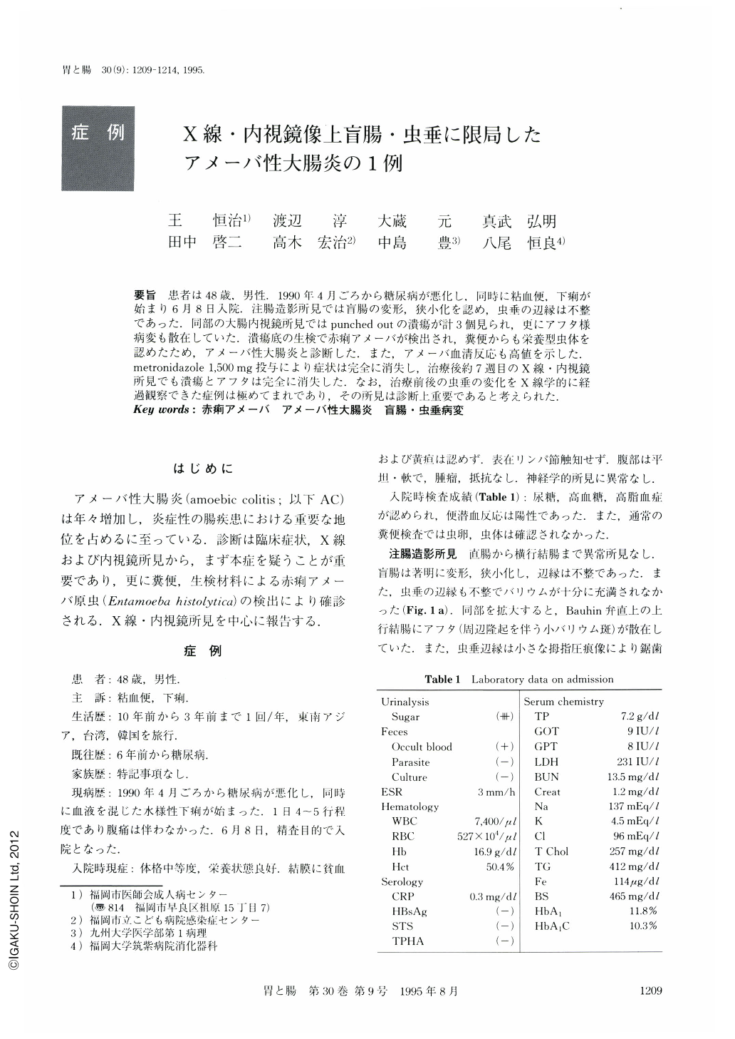
要旨 患者は48歳,男性.1990年4月ごろから糖尿病が悪化し.同時に粘血便.下痢が始まり6月8日入院.注腸造影所見では盲腸の変形,狭小化を認め,虫垂の辺縁は不整であった.同部の大腸内視鏡所見ではpunched outの潰瘍が計3個見られ,更にアフタ様病変も散在していた.潰瘍底の生検で赤痢アメーバが検出され,糞便からも栄養型虫体を認めたため,アメーバ性大腸炎と診断した.また,アメーバ血清反応も高値を示した.metronidazole 1,500mg投与により症状は完全に消失し,治療後約7週目のX線・内視鏡所見でも潰瘍とアフタは完全に消失した.なお,治療前後の虫垂の変化をX線学的に経過観察できた症例は極めてまれであり,その所見は診断上重要であると考えられた.
A 48-year-old man showed exacerbation of diabetes mellitus in April 1990, accompanied by mucous bloody stool and diarrhea. On June 8, 1990, he was admitted to our hospital. Barium enema study showed deformation and narrowing of the cecum and irregular margin of the appendix. Colonoscopic findings of this region revealed three punched out ulcers and scattered aphthoid erosions. Biopsy taken from the ulcer disclosed isolation of Entamoeba histolytica trophozites, and they were also found in feces. Based on these findings, the patient was diagnosed as having amoebic colitis. Serological test for Entamoeba histolytica was high in titer. Symptoms disappeared after treatment with metronidazole 1,500 mg per day. After seven weeks of therapy, radiographic and endoscopic findings no longer revealed ulcers and aphthoid erosions. This paper presents amoebic colitis restricted to the cecum and appendix, with special emphasis on its x-ray and endoscopic findings.

Copyright © 1995, Igaku-Shoin Ltd. All rights reserved.


