Japanese
English
- 有料閲覧
PPV購入案内
1,320円 (1,200円+税10%) こちらは、1109ページから1112ページの文献です。購入には医書.jp本会員登録が必要です。
会員登録完了後に改めて上記「購入サイトへ」を選択してください。
または「購入サイトへ」選択後に「購入ログイン」、「新規会員登録」の順に進んでください。
会員登録について詳しくはをご確認ください。
- Abstract 文献概要
- 1ページ目 Look Inside
- 参考文献 Reference
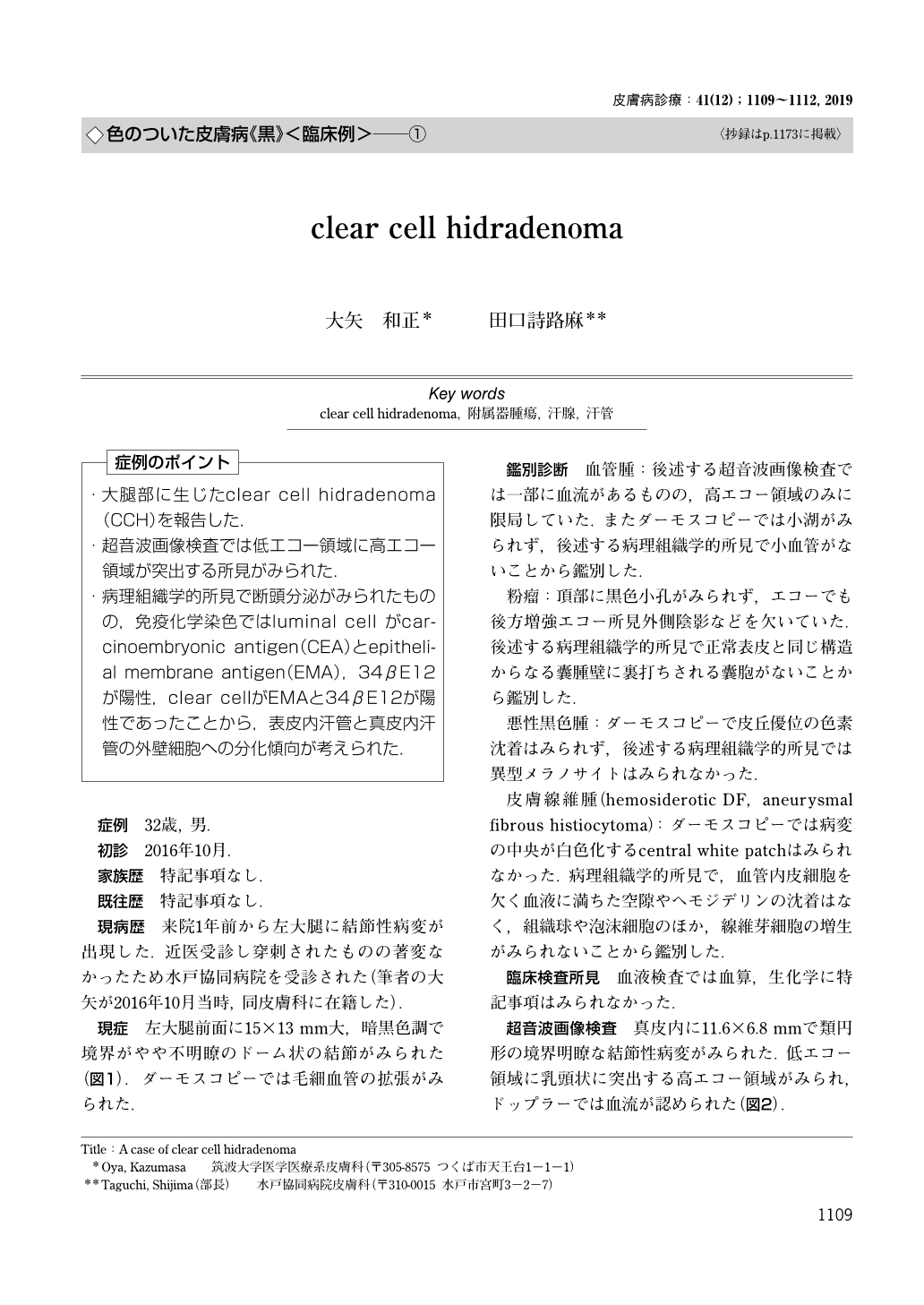
・大腿部に生じたclear cell hidradenoma(CCH)を報告した.
・超音波画像検査では低エコー領域に高エコー領域が突出する所見がみられた.
・病理組織学的所見で断頭分泌がみられたものの,免疫化学染色ではluminal cell がcarcinoembryonic antigen(CEA)とepithelial membrane antigen(EMA),34βE12が陽性,clear cellがEMAと34βE12が陽性であったことから,表皮内汗管と真皮内汗管の外壁細胞への分化傾向が考えられた.
(「症例のポイント」より)
A case of clear cell hidradenoma
Oya, Kazumasa1)Taguchi, Shijima2) 1)Department of Dermatology, Faculty of Medicine, University of Tsukuba 2)Dermatology, Mito Kyodo General Hospital
A 32-year-old male presented with a nodule on his left thigh. Physical examination revealed a 15×13-mm dark, dome-shaped nodule. Histopathological examination revealed that the mass consisted of solid and cystic areas. Clear cells and epidermoid cells were seen in the solid area. Duct-like structures lined by a layer of luminal cells were also present. Based on these findings, a diagnosis of clear cell hidradenoma was made. Immunohistochemical studies showed that the luminal cells were positive for carcinoembryonic antigen (CEA), epithelial membrane antigen (EMA) and 34βE12; and the clear cells were positive for EMA and 34βE12, which suggested the tumor had characteristics of acrosyringium with an outer layer consisting of dermal duct cells.
Copyright © 2019, KYOWA KIKAKU Ltd. All rights reserved.