Japanese
English
- 有料閲覧
PPV購入案内
1,320円 (1,200円+税10%) こちらは、949ページから952ページの文献です。購入には医書.jp本会員登録が必要です。
会員登録完了後に改めて上記「購入サイトへ」を選択してください。
または「購入サイトへ」選択後に「購入ログイン」、「新規会員登録」の順に進んでください。
会員登録について詳しくはをご確認ください。
- Abstract 文献概要
- 1ページ目 Look Inside
- 参考文献 Reference
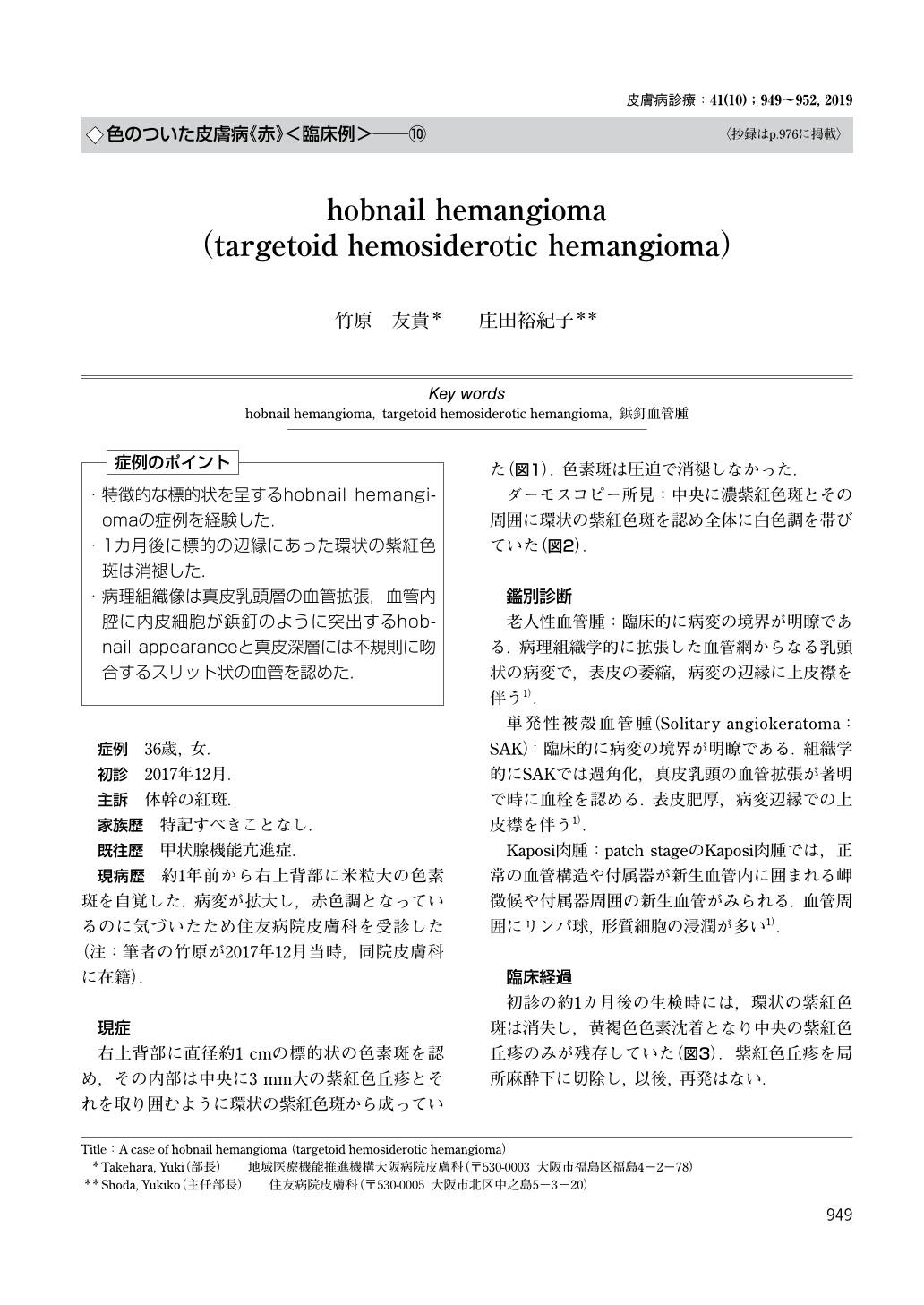
・特徴的な標的状を呈するhobnail hemangiomaの症例を経験した.
・1カ月後に標的の辺縁にあった環状の紫紅色斑は消褪した.
・病理組織像は真皮乳頭層の血管拡張,血管内腔に内皮細胞が鋲釘のように突出するhobnail appearanceと真皮深層には不規則に吻合するスリット状の血管を認めた.
(「症例のポイント」より)
A case of hobnail hemangioma (targetoid hemosiderotic hemangioma)
Takehara, Yuki1)Shoda, Yukiko2) 1)Department of Dermatology, Osaka Hospital, Japan Community Healthcare Organization(JCHO) 2)Department of Dermatology, Sumitomo Hospital
A 36-year-old women presented with a 10-mm red macule of one-year duration on her right upper back. The macule had a targetoid appearance, with an annular erythematous macule surrounding a 3-mm violaceous, slightly elevated papule. One month later, the annular macule had disappeared, and the residual papule was resected. A histological examination revealed a biphasic pattern with dilated superficial vessels, the endothelial cells of which were plump and projected intraluminally creating a “hobnail” appearance, and vascular slit-like space with flat endothelial cells in the deeper dermis. Based on the clinical and histological findings, we diagnosed the lesion as hobnail hemangioma.
Copyright © 2019, KYOWA KIKAKU Ltd. All rights reserved.