Japanese
English
- 有料閲覧
PPV購入案内
1,650円 (1,500円+税10%) こちらは、421ページから425ページの文献です。購入には医書.jp本会員登録が必要です。
会員登録完了後に改めて上記「購入サイトへ」を選択してください。
または「購入サイトへ」選択後に「購入ログイン」、「新規会員登録」の順に進んでください。
会員登録について詳しくはをご確認ください。
- Abstract 文献概要
- 1ページ目 Look Inside
- 参考文献 Reference
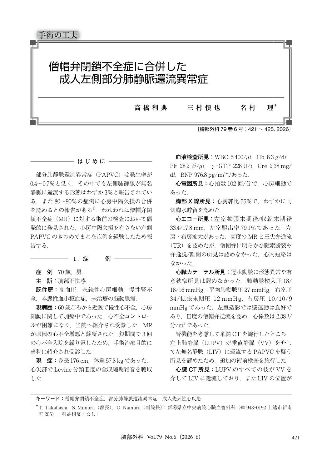
部分肺静脈還流異常症(PAPVC)は発生率が0.4~0.7%と低く,その中でも左側肺静脈が無名静脈に還流する形態はわずか3%と報告されている.また80~90%の症例に心房中隔欠損の合併を認めるとの報告がある1).われわれは僧帽弁閉鎖不全症(MR)に対する術前の検査において偶発的に発見された,心房中隔欠損を有さない左側PAPVCのきわめてまれな症例を経験したため報告する.
Partial anomalous pulmonary venous connection (PAPVC) is a rare congenital anomaly, and isolated left-sided PAPVC without atrial septal defect is extremely uncommon. We report a 70-year-old man with severe mitral regurgitation, atrial fibrillation, and chronic kidney disease, in whom preoperative imaging incidentally revealed left upper pulmonary vein drainage into the innominate vein via a vertical vein. Hemodynamic evaluation demonstrated a significant left-to-right shunt [pulmonary flow/systemic flow (Qp/Qs) =1.88]. Surgical treatment consisted of direct anastomosis of the vertical vein to the left atrial appendage, combined with mitral and tricuspid annuloplasty under cardiopulmonary bypass. The postoperative course was uneventful with stable hemodynamics. This case highlights the importance of careful preoperative imaging to detect rare anomalies such as PAPVC and the need for appropriate surgical planning when concomitant valvular disease is present.
© Nankodo Co., Ltd., 2026