Japanese
English
- 有料閲覧
PPV購入案内
1,320円 (1,200円+税10%) こちらは、431ページから438ページの文献です。購入には医書.jp本会員登録が必要です。
会員登録完了後に改めて上記「購入サイトへ」を選択してください。
または「購入サイトへ」選択後に「購入ログイン」、「新規会員登録」の順に進んでください。
会員登録について詳しくはをご確認ください。
- Abstract 文献概要
- 1ページ目 Look Inside
- 参考文献 Reference
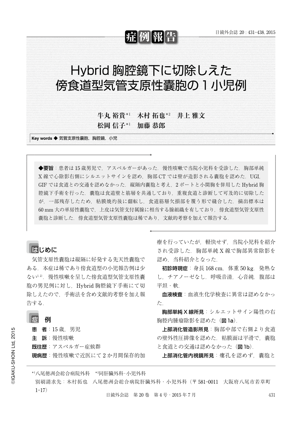
◆要旨:患者は15歳男児で,アスペルガーがあった.慢性咳嗽で当院小児科を受診した.胸部単純X線で心陰影右側にシルエットサインを認め,胸部CTでは壁が造影される囊胞を認めた.UGI,GIFでは食道との交通を認めなかった.縦隔内囊胞と考え,2ポートと小開胸を併用したHybrid胸腔鏡下手術を行った.囊胞は食道壁と筋層を共通しており,重複食道と診断して可及的に切除したが,一部残存したため,粘膜焼灼後に翻転し,食道筋層欠損部を覆う形で縫合した.摘出標本は60mm大の単房性囊胞で,上皮は気管支付属腺に相当する腺組織を有しており,傍食道型気管支原性囊胞と診断した.傍食道型気管支原性囊胞は稀であり,文献的考察を加えて報告する.
Bronchogenic cysts are a rare disease entity. We report a case of a 15-year-old male with a symptomatic bronchogenic cyst who underwent successful Video-assisted thoracoscopic excision. He was referred to our hospital because of a prolonged intermittent cough. Chest X-ray indicated a mediastinal mass. Computed to-mography(CT) and subsequent magnetic resonance imaging(MRI) demonstrated a 3.5×4.5×7.0 cm cystic lesion in the right posterior mediastinum without communicating esophagus. A video-assisted thoracoscopic approach was applied to excise the cyst. The medial aspect of the cyst was intimately involved with the muscular wall of the esophagus for several centimeters. A small portion of the cyst wall was left in place to avoid perforation of the esophagus. The remaining cyst wall was de-epithelialized. Intraoperative esophagoscopy revealed no esophageal injury. Post-operative course was uneventful. Histology achieved a diagnosis of bronchogenic cyst. The follow-up was well 18 mounths after the surgery. Bronchogenic cysts should be considered in the differential diagnoses for mediastinal masses at any age. Given their benign nature, thoracoscopy offers an excellent alternative to open thoracotomy for their removal particularly in children.
Copyright © 2015, JAPAN SOCIETY FOR ENDOSCOPIC SURGERY All rights reserved.