Japanese
English
- 有料閲覧
PPV購入案内
1,320円 (1,200円+税10%) こちらは、951ページから958ページの文献です。購入には医書.jp本会員登録が必要です。
会員登録完了後に改めて上記「購入サイトへ」を選択してください。
または「購入サイトへ」選択後に「購入ログイン」、「新規会員登録」の順に進んでください。
会員登録について詳しくはをご確認ください。
- Abstract 文献概要
- 1ページ目 Look Inside
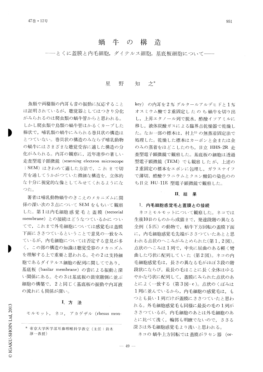
魚類や両棲類の内耳も音の振動に反応することは証明されているが,聴覚器としてはつきり分化がみられるのは爬虫類の蝸牛管からと思われる。しかし爬虫類や鳥類の蝸牛管はかるくカーブした棒状で,哺乳類の蝸牛にみられる巻貝状の構造はとつていない。巻貝状の構造のみならず哺乳動物の蝸牛にはさまざまな聴覚受容に適した構造の分化がみられる。内耳の観察に,近年進歩の著しい走査型電子顕微鏡(scanning electron microscope:SEM)はきわめて適した方法で,これまで切片を通してうかがつていた微細な構造を,立体的な十分に視覚的な像としてみせてくれるようになつた。
著者は哺乳動物蝸牛のきこえのメカニズムに関係の深い次の3点についてSEMをもちいて観察した。第1は内毛細胞感覚毛と蓋膜(tectorial membrane)との接続はどうなつているかについてで,これまで外毛細胞については感覚毛は蓋膜下面にささつているということで意見の一致をみているが,内毛細胞については否定する意見が多く,この部の構造の知識は聴覚受容のメカニズムを理解する上で重要と思われる。その2は支持細胞であるダイテルス細胞の配列に関してであり,基底板(basilar membrane)の音による振動と深い関係にある。その3は基底板の鼓室階側に並ぶ細胞の構築で,2と同じく基底板の振動や内耳液の流れにも関係が深い。
The fine structures of the cochlea of the rhesus monkey, the cat and the guinea pig were studied under a scanning electron microscope.
1. In the lower cochlea turns of the cat, impressions resulting from insertions of the inner sensory cell hair tips were observed on the under side of the tectorial membrane. These impressions were visible in the cat but not in the guinea pig. This aspect of the monkey cochlea was not-studied.
2. The phalangeal processes of the Deiters' cells of all animals examined were inclined toward the cochlea apex. Each process extended across the distince of 1 to 3 hair cells. The degree of inclination of the phalangeal processes of the 1st and 2nd outer hair cell rows were more pronounced than the processes associated with the 3rd outer hair cell row.
3. The mesothelial cells lining the scala tympani side of the basilar membrane were examined throughout the guinea pig cochlea. In the hook and lower 1/3 of the cochlea mesothelial cells were tightly packed together, whereas in the upper 2/3 of the cochlea these mesothelial cells had a spindle shape in contour with long thin processes on both ends of the cell body and these bodies were loosely arranged.
Copyright © 1975, Igaku-Shoin Ltd. All rights reserved.