Japanese
English
- 有料閲覧
PPV購入案内
1,320円 (1,200円+税10%) こちらは、527ページから531ページの文献です。購入には医書.jp本会員登録が必要です。
会員登録完了後に改めて上記「購入サイトへ」を選択してください。
または「購入サイトへ」選択後に「購入ログイン」、「新規会員登録」の順に進んでください。
会員登録について詳しくはをご確認ください。
- Abstract 文献概要
- 1ページ目 Look Inside
I.はじめに
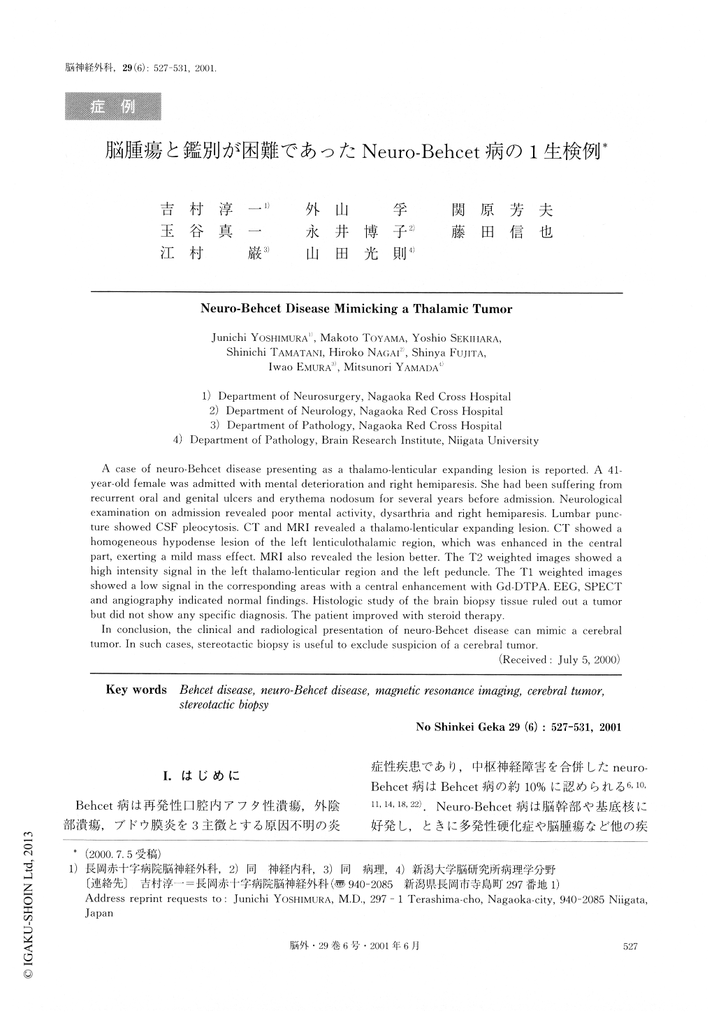
Behcet病は再発性口腔内アフタ性潰瘍,外陰部潰瘍,ブドウ膜炎を3主徴とする原因不明の炎症性疾患であり,中枢神経障害を合併したneuro-Behcet病はBehcet病の約10%に認められる6,10,11,14,18,22).Neuro-Behcet病は脳幹部や基底核に好発し,ときに多発性硬化症や脳腫瘍など他の疾患との鑑別が問題となる.
今回われわれは口腔内アフタ,外陰部潰瘍を繰り返し,若年の痴呆,右片麻痺にて発症した不全型neuro-Behcet病で術前脳腫瘍との鑑別が困難であった1生検例を経験したので報告する.
A case of neuro-Behcet disease presenting as a thalamo-lenticular expanding lesion is reported. A 41-year-old female was admitted with mental deterioration and right hemiparesis. She had been suffering fromrecurrent oral and genital ulcers and erythema nodosum for several years before admission. Neurologicalexamination on admission revealed poor mental activity, dysarthria and right hemiparesis. Lumbar punc-ture showed CSF pleocytosis. CT and MRI revealed a thalamo-lenticular expanding lesion. CT showed ahomogeneous hypodense lesion of the left lenticulothalamic region, which was enhanced in the centralpart, exerting a mild mass effect. MRI also revealed the lesion better. The T2 weighted images showed ahigh intensity signal in the left thalamo-lenticular region and the left peduncle. The Ti weighted imagesshowed a low signal in the corresponding areas with a central enhancement with Gd-DTPA. EEG, SPECTand angiography indicated normal findings. Histologic study of the brain biopsy tissue ruled out a tumorbut did not show any specific diagnosis. The patient improved with steroid therapy. In conclusion, the clinical and radiological presentation of neuro-Behcet disease can mimic a cerebraltumor. In such cases, stereotactic biopsy is useful to exclude suspicion of a cerebral tumor.
Copyright © 2001, Igaku-Shoin Ltd. All rights reserved.