Japanese
English
- 有料閲覧
PPV購入案内
1,320円 (1,200円+税10%) こちらは、259ページから264ページの文献です。購入には医書.jp本会員登録が必要です。
会員登録完了後に改めて上記「購入サイトへ」を選択してください。
または「購入サイトへ」選択後に「購入ログイン」、「新規会員登録」の順に進んでください。
会員登録について詳しくはをご確認ください。
- Abstract 文献概要
- 1ページ目 Look Inside
I.はじめに
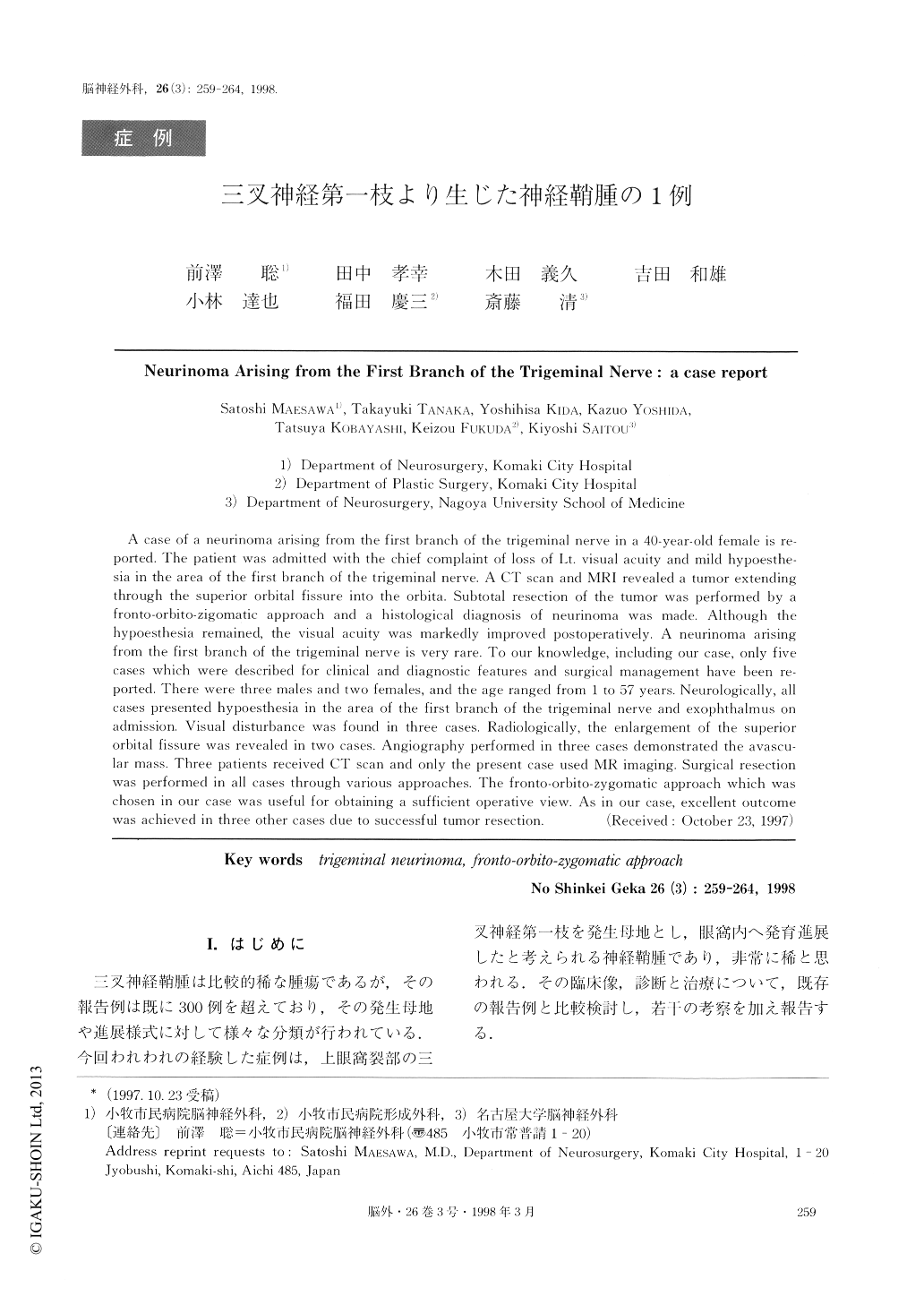
三叉神経鞘腫は比較的稀な腫瘍であるが,その報告例は既に300例を超えており,その発生母地や進展様式に対して様々な分類が行われている.今回われわれの経験した症例は,上眼窩裂部の三叉神経第一枝を発生母地とし,眼窩内へ発育進展したと考えられる神経鞘腫であり,非常に稀と思われる.その臨床像,診断と治療について,既存の報告例と比較検討し,若干の考察を加え報告する.
A case of a neurinoma arising from the first branch of the trigeminal nerve in a 40-year-old female is re-ported, The patient was admitted with the chief complaint of loss of Lt. visual acuity and mild hypoesthe-sia in the area of the first branch of the trigeminal nerve. A CT scan and MRI revealed a tumor extendingthrough the superior orbitai fissure into the orbita. Subtotal resection of the tumor was performed by afronto-orbito-zigomatic approach and a histoiogical diagnosis of neurinoma was made. Although thehypoesthesia remained, the visual acuity was markedly improved postoperatively. A neurinoma arisingfrom the first branch of the trigeminal nerve is very rare. To our knowledge, including our case, only fivecases which were described for clinical and diagnostic features and surgical management have been re-ported. There were three males and two females, and the age ranged from 1 to 57 years. Neurologically, allcases presented hypoesthesia in the area of the first branch of the trigeminal nerve and exophthalmus onadmission. Visual disturbance was found in three cases. Radiologically, the enlargement of the superiororbital fissure was revealed in two cases, Angiography performed in three cases demonstrated the avascu-lar mass. Three patients received CT scan and only the present case used MR imaging. Surgical resectionwas performed in all cases through various approaches. The fronto-orbito-zygomatic approach which waschosen in our case was useful for obtaining a sufficient operative view. As in our case, excellent outcomewas achieved in three other cases due to successful tumor resection.
Copyright © 1998, Igaku-Shoin Ltd. All rights reserved.