Japanese
English
- 有料閲覧
PPV購入案内
1,320円 (1,200円+税10%) こちらは、837ページから841ページの文献です。購入には医書.jp本会員登録が必要です。
会員登録完了後に改めて上記「購入サイトへ」を選択してください。
または「購入サイトへ」選択後に「購入ログイン」、「新規会員登録」の順に進んでください。
会員登録について詳しくはをご確認ください。
- Abstract 文献概要
- 1ページ目 Look Inside
I.はじめに
黄色腫は,一般に高脂血症や糖尿病などの代謝異常症に合併し,皮膚や軟部組織に発生する疾患で,骨原発のものは稀であり,とりわけ頭蓋骨に発生したものの報告はきわめて少ない.われわれが経験したxanthofibromaに関し,若干の文献的考察を加え報告する.
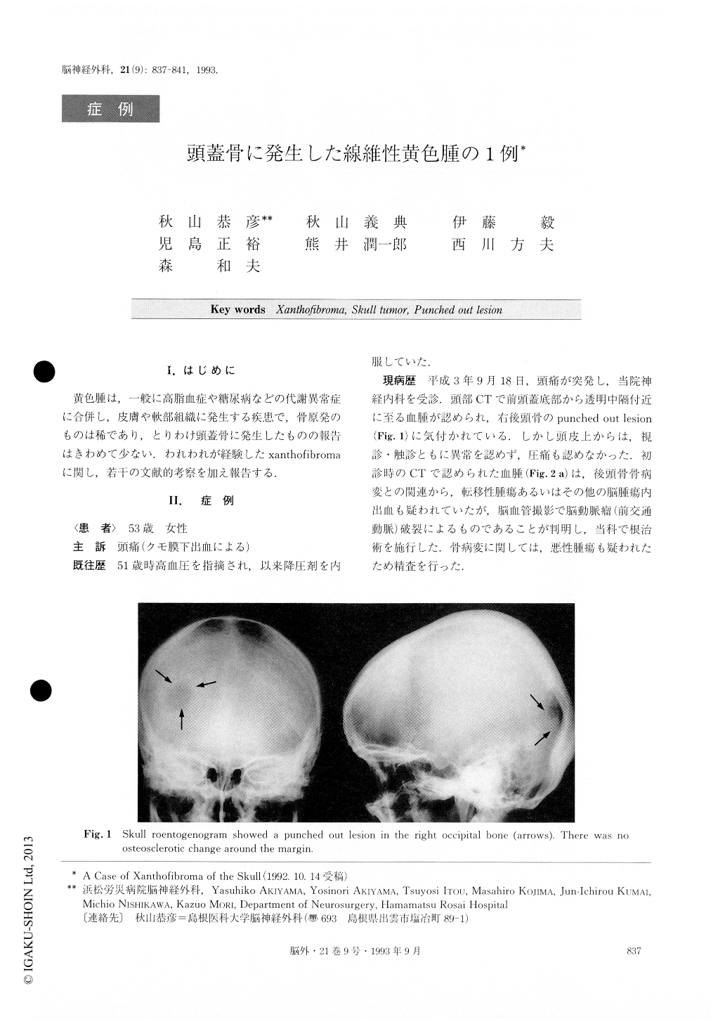
The authors describe a case of xanthofibroma of the skull. A 53-year-old female was admitted to our hospital in September, 1991, with subarachnoid hemorrhage dueto a ruptured aneurysm of the anterior communicating artery. The roentogenogram of her skull incidentally rev-ealed the presence of a radiolucent (a punched out) le-sion of about 20×25mm in the right occipital bone.
On computed tomography (CT), the mass was seen to be mainly localized in the diploe, and the outer table of the skull was thinned. On both T1 and T2 weighted magnetic resonance image (MRI), the mass showed a high intensity signal equivalent to that found in adipose tissue.
The bony tumor was totally removed. Histology re-vealed a collection of foamy cells, benign fibrous tissues and so on which led to a diagnosis of xanthofibroma of the skull without hyperlipidemia.
Xanthofibroma of the skull is extremely rare. To our knowledge, including our case, only three cases have been reported.
Copyright © 1993, Igaku-Shoin Ltd. All rights reserved.