Japanese
English
- 有料閲覧
PPV購入案内
1,320円 (1,200円+税10%) こちらは、288ページから297ページの文献です。購入には医書.jp本会員登録が必要です。
会員登録完了後に改めて上記「購入サイトへ」を選択してください。
または「購入サイトへ」選択後に「購入ログイン」、「新規会員登録」の順に進んでください。
会員登録について詳しくはをご確認ください。
- Abstract 文献概要
- 1ページ目 Look Inside
- 参考文献 Reference
Point
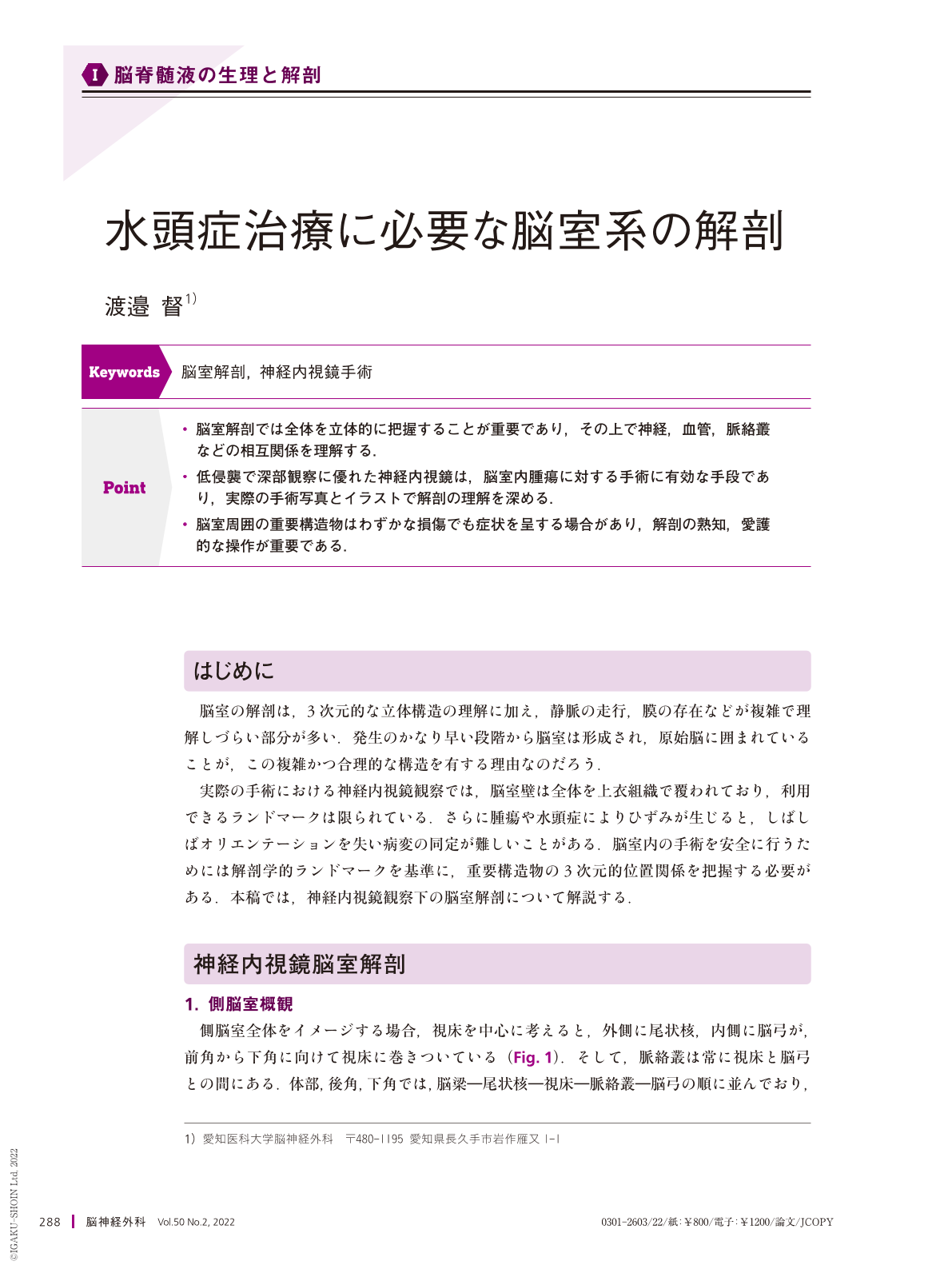
・脳室解剖では全体を立体的に把握することが重要であり,その上で神経,血管,脈絡叢などの相互関係を理解する.
・低侵襲で深部観察に優れた神経内視鏡は,脳室内腫瘍に対する手術に有効な手段であり,実際の手術写真とイラストで解剖の理解を深める.
・脳室周囲の重要構造物はわずかな損傷でも症状を呈する場合があり,解剖の熟知,愛護的な操作が重要である.
Minimally invasive endoscopes with excellent deep observation capabilities are effective means for surgery for intraventricular lesions. The ventricles have a complex three-dimensionally structure, and important structures around the ventricles may causes symptoms even with slight damage. Therefore, familiarity with the ventricular anatomy and protective manipulations are important. During endoscopic observations in actual surgery, the ventricular wall is entirely covered with the ependymal tissue, limiting the visibility of landmarks. Furthermore, when distorted by the tumor, the anatomical orientation may be lost, and identifying the lesion can be difficult. In this chapter, we describe the ventricular anatomy under endoscopic observation using actual photographs and illustrations based on anatomical landmarks, in order to safely perform intraventricular surgery.
Copyright © 2022, Igaku-Shoin Ltd. All rights reserved.