Japanese
English
- 有料閲覧
PPV購入案内
1,320円 (1,200円+税10%) こちらは、547ページから552ページの文献です。購入には医書.jp本会員登録が必要です。
会員登録完了後に改めて上記「購入サイトへ」を選択してください。
または「購入サイトへ」選択後に「購入ログイン」、「新規会員登録」の順に進んでください。
会員登録について詳しくはをご確認ください。
- Abstract 文献概要
- 1ページ目 Look Inside
- 参考文献 Reference
Ⅰ.はじめに
前頭蓋窩硬膜動静脈瘻(anterior cranial fossa dural arteriovenous fistula:ACF-DAVF)は全DAVF中6%程度の頻度の稀な疾患であるが,そのうち61〜91%は脳出血を契機に発見されると報告されており1,5,9),てんかん発作で発見される症例はさらに稀である.一般的に,DAVFは発生部位・症状が多様であり,神経学的症候のみで積極的にこの疾患を疑うことが困難な場合もあり,診断に苦慮することも珍しくはない.画像診断のgold standardは脳血管造影(digital subtraction angiography:DSA)検査であるが,侵襲度が高い検査であり,強く疑われる症例にのみ適応されるべきと考えられる.Arterial spin-labeling imaging(ASL画像)は,造影剤を用いずに脳組織灌流状態を撮像できるMRI検査法の1つであり,脳血管障害や脳腫瘍などの血流評価に臨床応用されている.
今回,特徴的なASL画像所見に基づき診断に至った,てんかん発症のACF-DAVFの症例を経験したので報告する.
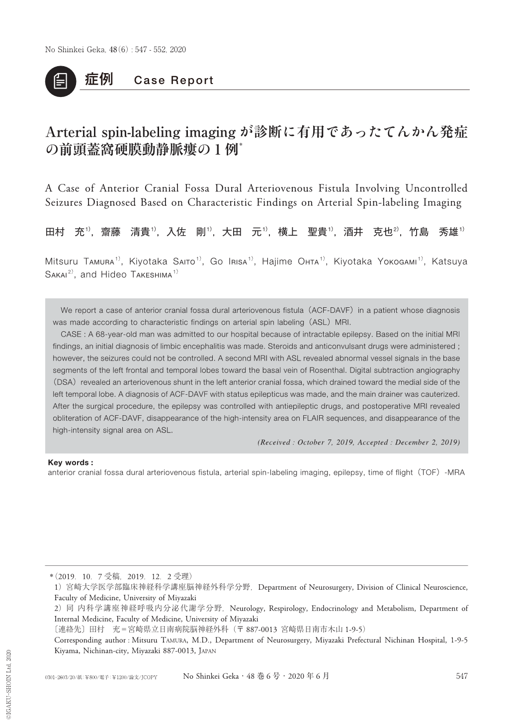
We report a case of anterior cranial fossa dural arteriovenous fistula(ACF-DAVF)in a patient whose diagnosis was made according to characteristic findings on arterial spin labeling(ASL)MRI.
CASE:A 68-year-old man was admitted to our hospital because of intractable epilepsy. Based on the initial MRI findings, an initial diagnosis of limbic encephalitis was made. Steroids and anticonvulsant drugs were administered;however, the seizures could not be controlled. A second MRI with ASL revealed abnormal vessel signals in the base segments of the left frontal and temporal lobes toward the basal vein of Rosenthal. Digital subtraction angiography(DSA)revealed an arteriovenous shunt in the left anterior cranial fossa, which drained toward the medial side of the left temporal lobe. A diagnosis of ACF-DAVF with status epilepticus was made, and the main drainer was cauterized. After the surgical procedure, the epilepsy was controlled with antiepileptic drugs, and postoperative MRI revealed obliteration of ACF-DAVF, disappearance of the high-intensity area on FLAIR sequences, and disappearance of the high-intensity signal area on ASL.
Copyright © 2020, Igaku-Shoin Ltd. All rights reserved.