Japanese
English
- 有料閲覧
PPV購入案内
1,320円 (1,200円+税10%) こちらは、413ページから422ページの文献です。購入には医書.jp本会員登録が必要です。
会員登録完了後に改めて上記「購入サイトへ」を選択してください。
または「購入サイトへ」選択後に「購入ログイン」、「新規会員登録」の順に進んでください。
会員登録について詳しくはをご確認ください。
- Abstract 文献概要
- 1ページ目 Look Inside
- 参考文献 Reference
Ⅰ.はじめに
非外傷性急性硬膜下血腫(acute subdural hematoma:ASDH)を伴って発症する髄膜腫2,6)は散見されるが,半球間裂主体に発生した報告例は少ない10,12,16,17).今回われわれは,腫瘍内出血と半球間裂ASDHにより発症した大脳鎌髄膜腫の稀な1例を経験した.本例の出血機序や初期診療における問題点などについて,文献的考察を加えて報告する.
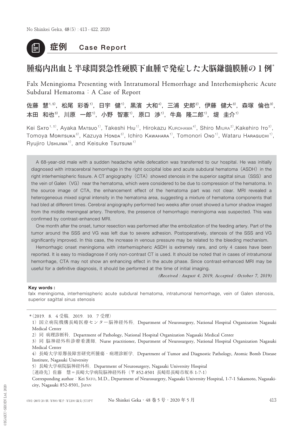
A 68-year-old male with a sudden headache while defecation was transferred to our hospital. He was initially diagnosed with intracerebral hemorrhage in the right occipital lobe and acute subdural hematoma(ASDH)in the right interhemispheric fissure. A CT angiography(CTA)showed stenosis in the superior sagittal sinus(SSS)and the vein of Galen(VG)near the hematoma, which were considered to be due to compression of the hematoma. In the source image of CTA, the enhancement effect of the hematoma part was not clear. MRI revealed a heterogeneous mixed signal intensity in the hematoma area, suggesting a mixture of hematoma components that had bled at different times. Cerebral angiography performed two weeks after onset showed a tumor shadow imaged from the middle meningeal artery. Therefore, the presence of hemorrhagic meningioma was suspected. This was confirmed by contrast-enhanced MRI.
One month after the onset, tumor resection was performed after the embolization of the feeding artery. Part of the tumor around the SSS and VG was left due to severe adhesion. Postoperatively, stenosis of the SSS and VG significantly improved. In this case, the increase in venous pressure may be related to the bleeding mechanism.
Hemorrhagic onset meningioma with interhemispheric ASDH is extremely rare, and only 4 cases have been reported. It is easy to misdiagnose if only non-contrast CT is used. It should be noted that in cases of intratumoral hemorrhage, CTA may not show an enhancing effect in the acute phase. Since contrast-enhanced MRI may be useful for a definitive diagnosis, it should be performed at the time of initial imaging.
Copyright © 2020, Igaku-Shoin Ltd. All rights reserved.