Japanese
English
- 有料閲覧
PPV購入案内
1,320円 (1,200円+税10%) こちらは、1073ページから1079ページの文献です。購入には医書.jp本会員登録が必要です。
会員登録完了後に改めて上記「購入サイトへ」を選択してください。
または「購入サイトへ」選択後に「購入ログイン」、「新規会員登録」の順に進んでください。
会員登録について詳しくはをご確認ください。
- Abstract 文献概要
- 1ページ目 Look Inside
- 参考文献 Reference
Ⅰ.はじめに
脳腫瘍で腫瘍内出血を来す頻度は0.6〜14.6%とされ,出血を来しやすいのは一般的に転移性脳腫瘍や膠芽腫などの悪性脳腫瘍である21).腫瘍内出血の頻度は,転移性脳腫瘍では2.9〜14%で,原疾患として,肺癌,悪性黒色腫,絨毛癌,甲状腺癌が多い16).また,原発性脳腫瘍である神経膠腫では3.7〜7.2%の頻度であり,膠芽腫の頻度が最も高く,low grade glioma(LGG)は1%未満と稀である1,5,10,15).今回われわれは,腫瘍内出血で発症し,脳血管撮影において動静脈短絡を呈したdiffuse astrocytomaの1例を経験したので報告する.
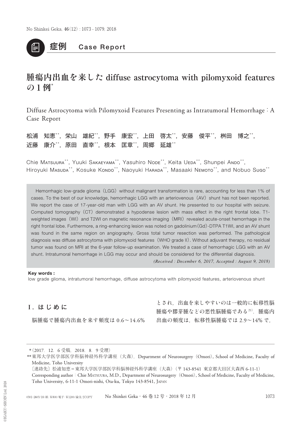
Hemorrhagic low-grade glioma(LGG)without malignant transformation is rare, accounting for less than 1% of cases. To the best of our knowledge, hemorrhagic LGG with an arteriovenous(AV)shunt has not been reported. We report the case of 17-year-old man with LGG with an AV shunt. He presented to our hospital with seizure. Computed tomography(CT)demonstrated a hypodense lesion with mass effect in the right frontal lobe. T1-weighted images(WI)and T2WI on magnetic resonance imaging(MRI)revealed acute-onset hemorrhage in the right frontal lobe. Furthermore, a ring-enhancing lesion was noted on gadolinium(Gd)-DTPA T1WI, and an AV shunt was found in the same region on angiography. Gross total tumor resection was performed. The pathological diagnosis was diffuse astrocytoma with pilomyxoid features(WHO grade Ⅱ). Without adjuvant therapy, no residual tumor was found on MRI at the 6-year follow-up examination. We treated a case of hemorrhagic LGG with an AV shunt. Intratumoral hemorrhage in LGG may occur and should be considered for the differential diagnosis.
Copyright © 2018, Igaku-Shoin Ltd. All rights reserved.