Japanese
English
- 有料閲覧
PPV購入案内
1,320円 (1,200円+税10%) こちらは、1071ページから1077ページの文献です。購入には医書.jp本会員登録が必要です。
会員登録完了後に改めて上記「購入サイトへ」を選択してください。
または「購入サイトへ」選択後に「購入ログイン」、「新規会員登録」の順に進んでください。
会員登録について詳しくはをご確認ください。
- Abstract 文献概要
- 1ページ目 Look Inside
- 参考文献 Reference
Ⅰ.はじめに
眼科以外での手術後に,失明などの視力障害の発症を0.05~1%に認めることが以前から報告されており,その原因として,直接的な視神経損傷以外に,虚血性視神経症,網膜中心動脈閉塞症,網膜中心静脈閉塞症,皮質盲などが考えられている2,3).両側前頭開頭や前頭側頭開頭後においても1%以下の頻度ではあるが失明を来す報告があり,いったん発生するとその予後は極めて不良とされている2,3).開頭手術時に失明を来す要因としては,翻転された皮膚弁により眼球に圧迫が加わることで眼球内圧が亢進し,網膜や視神経を栄養している網膜中心動脈や短後毛様体動脈の循環不全を来すことが一因であると考えられている2).
当施設でも,前交通動脈瘤に対する両側前頭開頭でのbasal interhemispheric approachによる手術後に,片側の視力障害を呈した例を経験した.そこで2008年より開頭術時にFlexiForceセンサー(A201-1,ニッタ,大阪)による眼圧モニタリングを開始した.既に本センサーが眼球への過度の圧迫を防止する上で有効であることを報告したが4),今回症例を蓄積し新たな知見を得たので,その結果を報告する.
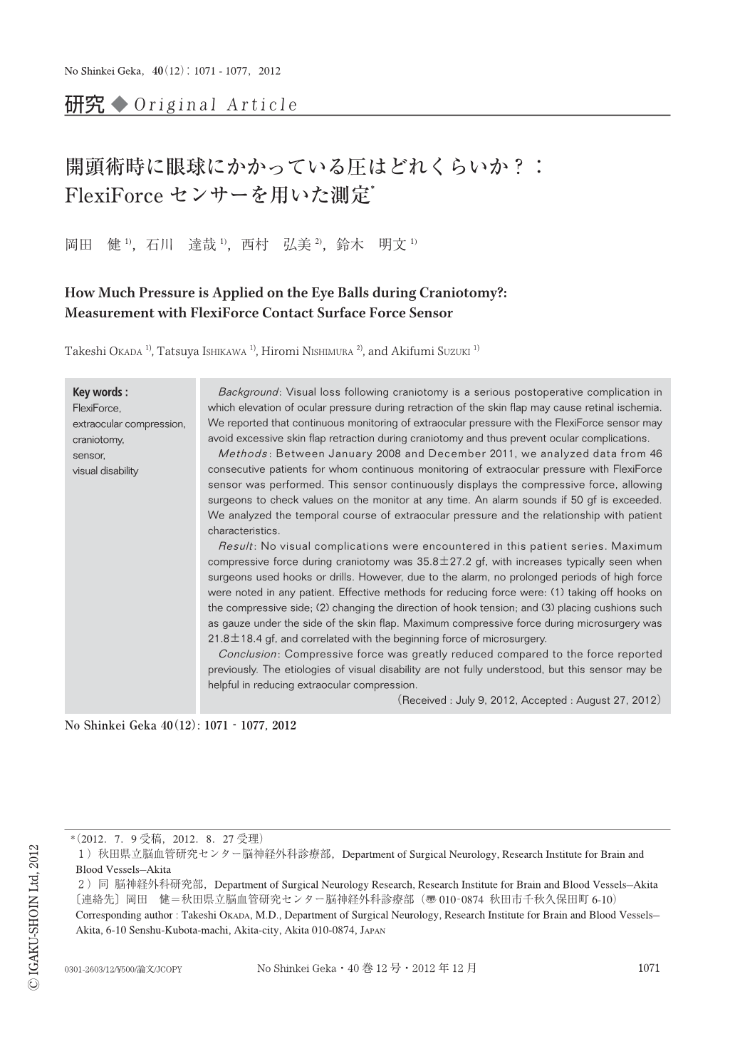
Background: Visual loss following craniotomy is a serious postoperative complication in which elevation of ocular pressure during retraction of the skin flap may cause retinal ischemia. We reported that continuous monitoring of extraocular pressure with the FlexiForce sensor may avoid excessive skin flap retraction during craniotomy and thus prevent ocular complications.
Methods: Between January 2008 and December 2011, we analyzed data from 46 consecutive patients for whom continuous monitoring of extraocular pressure with FlexiForce sensor was performed. This sensor continuously displays the compressive force, allowing surgeons to check values on the monitor at any time. An alarm sounds if 50 gf is exceeded. We analyzed the temporal course of extraocular pressure and the relationship with patient characteristics.
Result: No visual complications were encountered in this patient series. Maximum compressive force during craniotomy was 35.8±27.2 gf, with increases typically seen when surgeons used hooks or drills. However, due to the alarm, no prolonged periods of high force were noted in any patient. Effective methods for reducing force were: (1) taking off hooks on the compressive side; (2) changing the direction of hook tension; and (3) placing cushions such as gauze under the side of the skin flap. Maximum compressive force during microsurgery was 21.8±18.4 gf, and correlated with the beginning force of microsurgery.
Conclusion: Compressive force was greatly reduced compared to the force reported previously. The etiologies of visual disability are not fully understood, but this sensor may be helpful in reducing extraocular compression.
Copyright © 2012, Igaku-Shoin Ltd. All rights reserved.