Japanese
English
- 有料閲覧
PPV購入案内
1,320円 (1,200円+税10%) こちらは、877ページから881ページの文献です。購入には医書.jp本会員登録が必要です。
会員登録完了後に改めて上記「購入サイトへ」を選択してください。
または「購入サイトへ」選択後に「購入ログイン」、「新規会員登録」の順に進んでください。
会員登録について詳しくはをご確認ください。
- Abstract 文献概要
- 1ページ目 Look Inside
- 参考文献 Reference
Ⅰ.はじめに
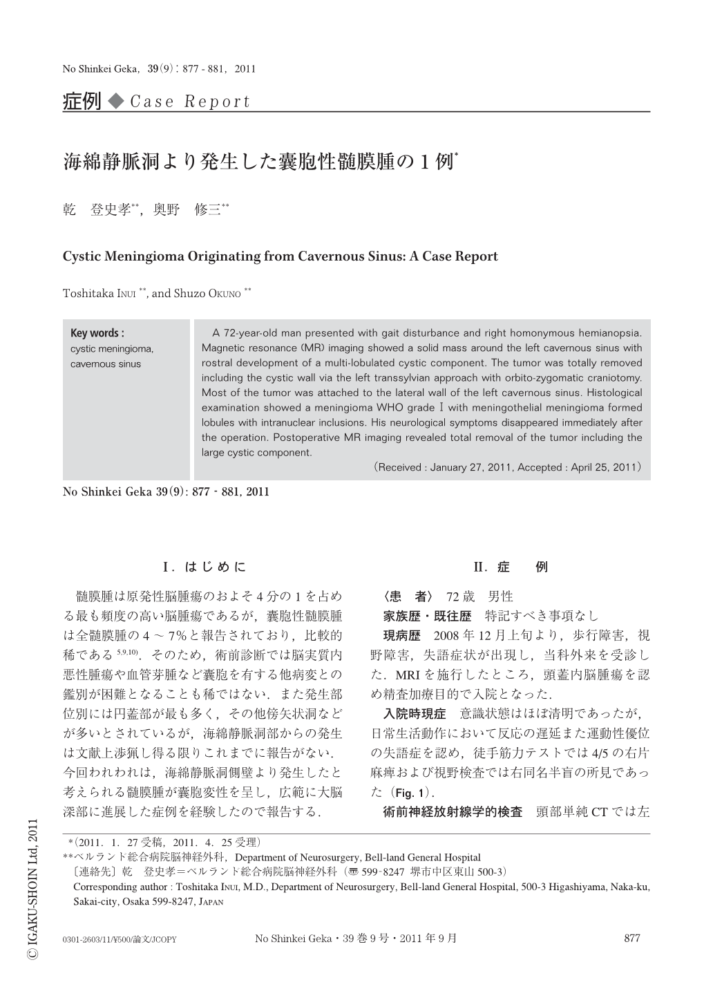
髄膜腫は原発性脳腫瘍のおよそ4分の1を占める最も頻度の高い脳腫瘍であるが,囊胞性髄膜腫は全髄膜腫の4~7%と報告されており,比較的稀である5,9,10).そのため,術前診断では脳実質内悪性腫瘍や血管芽腫など囊胞を有する他病変との鑑別が困難となることも稀ではない.また発生部位別には円蓋部が最も多く,その他傍矢状洞などが多いとされているが,海綿静脈洞部からの発生は文献上渉猟し得る限りこれまでに報告がない.今回われわれは,海綿静脈洞側壁より発生したと考えられる髄膜腫が囊胞変性を呈し,広範に大脳深部に進展した症例を経験したので報告する.
A 72-year-old man presented with gait disturbance and right homonymous hemianopsia. Magnetic resonance (MR) imaging showed a solid mass around the left cavernous sinus with rostral development of a multi-lobulated cystic component. The tumor was totally removed including the cystic wall via the left transsylvian approach with orbito-zygomatic craniotomy. Most of the tumor was attached to the lateral wall of the left cavernous sinus. Histological examination showed a meningioma WHO gradeⅠwith meningothelial meningioma formed lobules with intranuclear inclusions. His neurological symptoms disappeared immediately after the operation. Postoperative MR imaging revealed total removal of the tumor including the large cystic component.
Copyright © 2011, Igaku-Shoin Ltd. All rights reserved.