Japanese
English
- 有料閲覧
PPV購入案内
1,320円 (1,200円+税10%) こちらは、53ページから59ページの文献です。購入には医書.jp本会員登録が必要です。
会員登録完了後に改めて上記「購入サイトへ」を選択してください。
または「購入サイトへ」選択後に「購入ログイン」、「新規会員登録」の順に進んでください。
会員登録について詳しくはをご確認ください。
- Abstract 文献概要
- 1ページ目 Look Inside
- 参考文献 Reference
Ⅰ.はじめに
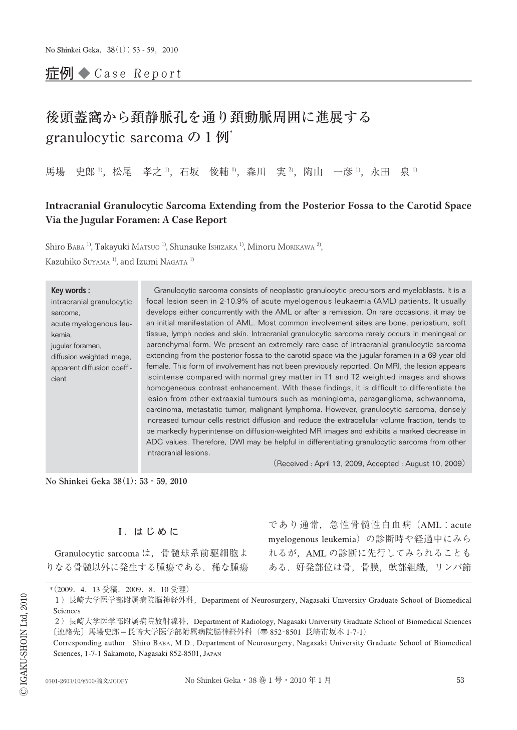
Granulocytic sarcomaは,骨髄球系前駆細胞よりなる骨髄以外に発生する腫瘍である.稀な腫瘍であり通常,急性骨髄性白血病(AML:acute myelogenous leukemia)の診断時や経過中にみられるが,AMLの診断に先行してみられることもある.好発部位は骨,骨膜,軟部組織,リンパ節および皮膚であり,頭蓋内での発生は稀である.今回われわれは,後頭蓋窩から頚静脈孔を通り頚動脈周囲に進展した極めて稀な症例を経験し,またMRI拡散強調画像(DWI:diffusion weighted image)が術前診断に有用であったため,文献的考察を加えて報告する.
Granulocytic sarcoma consists of neoplastic granulocytic precursors and myeloblasts. It is a focal lesion seen in 2-10.9% of acute myelogenous leukaemia (AML) patients. It usually develops either concurrently with the AML or after a remission. On rare occasions,it may be an initial manifestation of AML. Most common involvement sites are bone,periostium,soft tissue,lymph nodes and skin. Intracranial granulocytic sarcoma rarely occurs in meningeal or parenchymal form. We present an extremely rare case of intracranial granulocytic sarcoma extending from the posterior fossa to the carotid space via the jugular foramen in a 69 year old female. This form of involvement has not been previously reported. On MRI,the lesion appears isointense compared with normal grey matter in T1 and T2 weighted images and shows homogeneous contrast enhancement. With these findings,it is difficult to differentiate the lesion from other extraaxial tumours such as meningioma,paraganglioma,schwannoma,carcinoma,metastatic tumor,malignant lymphoma. However,granulocytic sarcoma,densely increased tumour cells restrict diffusion and reduce the extracellular volume fraction,tends to be markedly hyperintense on diffusion-weighted MR images and exhibits a marked decrease in ADC values. Therefore,DWI may be helpful in differentiating granulocytic sarcoma from other intracranial lesions.
Copyright © 2010, Igaku-Shoin Ltd. All rights reserved.