Japanese
English
- 有料閲覧
PPV購入案内
1,320円 (1,200円+税10%) こちらは、49ページから54ページの文献です。購入には医書.jp本会員登録が必要です。
会員登録完了後に改めて上記「購入サイトへ」を選択してください。
または「購入サイトへ」選択後に「購入ログイン」、「新規会員登録」の順に進んでください。
会員登録について詳しくはをご確認ください。
- Abstract 文献概要
- 1ページ目 Look Inside
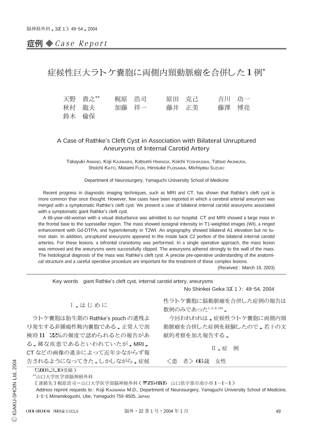
Ⅰ.はじめに
ラトケ囊胞は胎生期のRathke's pouchの遺残より発生する非腫瘍性鞍内囊胞である.正常人で剖検時11~22%の頻度で認められるとの報告がある.稀な疾患であるといわれていたが,MRI,CTなどの画像の進歩によって近年少なからず報告されるようになってきた.しかしながら,症候性ラトケ囊胞に脳動脈瘤を合併した症例の報告は数例のみであった1,5,9,10).
今回われわれは,症候性ラトケ囊胞に両側内頸動脈瘤を合併した症例を経験したので,若干の文献的考察を加え報告する.
Recent progress in diagnostic imaging techniques,such as MRI and CT,has shown that Rathke's cleft cyst is more common than once thought. However,few cases have been reported in which a cerebral arterial aneurysm was merged with a symptomatic Rathke's cleft cyst. We present a case of bilateral internal carotid aneurysms associated with a symptomatic giant Rathke's cleft cyst.
A 66-year-old-woman with a visual disturbance was admitted to our hospital. CT and MRI showed a large mass in the frontal base to the suprasellar region. The mass showed isosignal intensity in T1-weighted images (WI),a ringed enhancement with Gd-DTPA,and hyperintensity in T2WI. An angiography showed bilateral A1 elevation but no tumor stain. In addition,unruptured aneurysms appeared in the inside back C2 portion of the bilateral internal carotid arteries. For these lesions,a bifrontal craniotomy was performed. In a single operative approach,the mass lesion was removed and the aneurysms were successfully clipped. The aneurysms adhered strongly to the wall of the mass. The histological diagnosis of the mass was Rathke's cleft cyst. A precise pre-operative understanding of the anatomical structure and a careful operative procedure are important for the treatment of these complex lesions.
Copyright © 2004, Igaku-Shoin Ltd. All rights reserved.