Japanese
English
- 有料閲覧
PPV購入案内
1,320円 (1,200円+税10%) こちらは、851ページから856ページの文献です。購入には医書.jp本会員登録が必要です。
会員登録完了後に改めて上記「購入サイトへ」を選択してください。
または「購入サイトへ」選択後に「購入ログイン」、「新規会員登録」の順に進んでください。
会員登録について詳しくはをご確認ください。
- Abstract 文献概要
- 1ページ目 Look Inside
- 参考文献 Reference
Ⅰ.は じ め に
慢性被膜化脳内血腫(encapsulized intracranial hematoma; 以下EIH)は脳内血腫の特殊な一形態であり,過去の報告例も少なく詳細な病態はいまだ不明である.また,画像所見による確定診断は困難で腫瘍性病変と診断されることが少なくない.今回われわれは術前画像診断で悪性脳腫瘍と診断したが,病理所見でEIHと診断された稀な1例を経験したので,文献的考察を加え報告する.
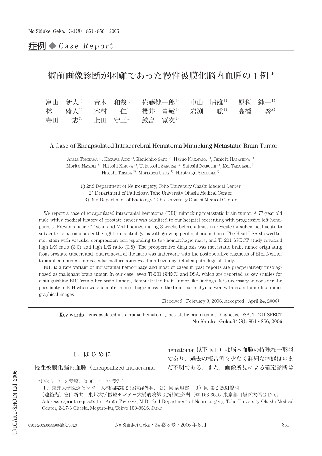
We report a case of encapsulated intracranial hematoma (EIH) mimicking metastatic brain tumor. A 77-year old male with a medical history of prostate cancer was admitted to our hospital presenting with progressive left hemiparesis. Previous head CT scan and MRI findings during 3 weeks before admission revealed a subcortical acute to subacute hematoma under the right precentral gyrus with growing perifocal brainedema. The Head DSA showed tumor-stain with vascular compression corresponding to the hemorrhagic mass,and Tl-201 SPECT study revealed high L/N ratio (3.0) and high L/E ratio (0.8). The preoperative diagnosis was metastatic brain tumor originating from prostate cancer,and total removal of the mass was undergone with the postoperative diagnosis of EIH. Neither tumoral component nor vascular malformation was found even by detailed pathological study.
EIH is a rare variant of intracranial hemorrhage and most of cases in past reports are preoperatively misdiagnosed as malignant brain tumor. In our case,even Tl-201 SPECT and DSA,which are reported as key studies for distinguishing EIH from other brain tumors,demonstrated brain tumor-like findings. It is necessary to consider the possibility of EIH when we encounter hemorrhagic mass in the brain parenchyma even with brain tumor-like radiographical images.
Copyright © 2006, Igaku-Shoin Ltd. All rights reserved.