Japanese
English
- 有料閲覧
PPV購入案内
1,320円 (1,200円+税10%) こちらは、725ページから730ページの文献です。購入には医書.jp本会員登録が必要です。
会員登録完了後に改めて上記「購入サイトへ」を選択してください。
または「購入サイトへ」選択後に「購入ログイン」、「新規会員登録」の順に進んでください。
会員登録について詳しくはをご確認ください。
- Abstract 文献概要
- 1ページ目 Look Inside
- 参考文献 Reference
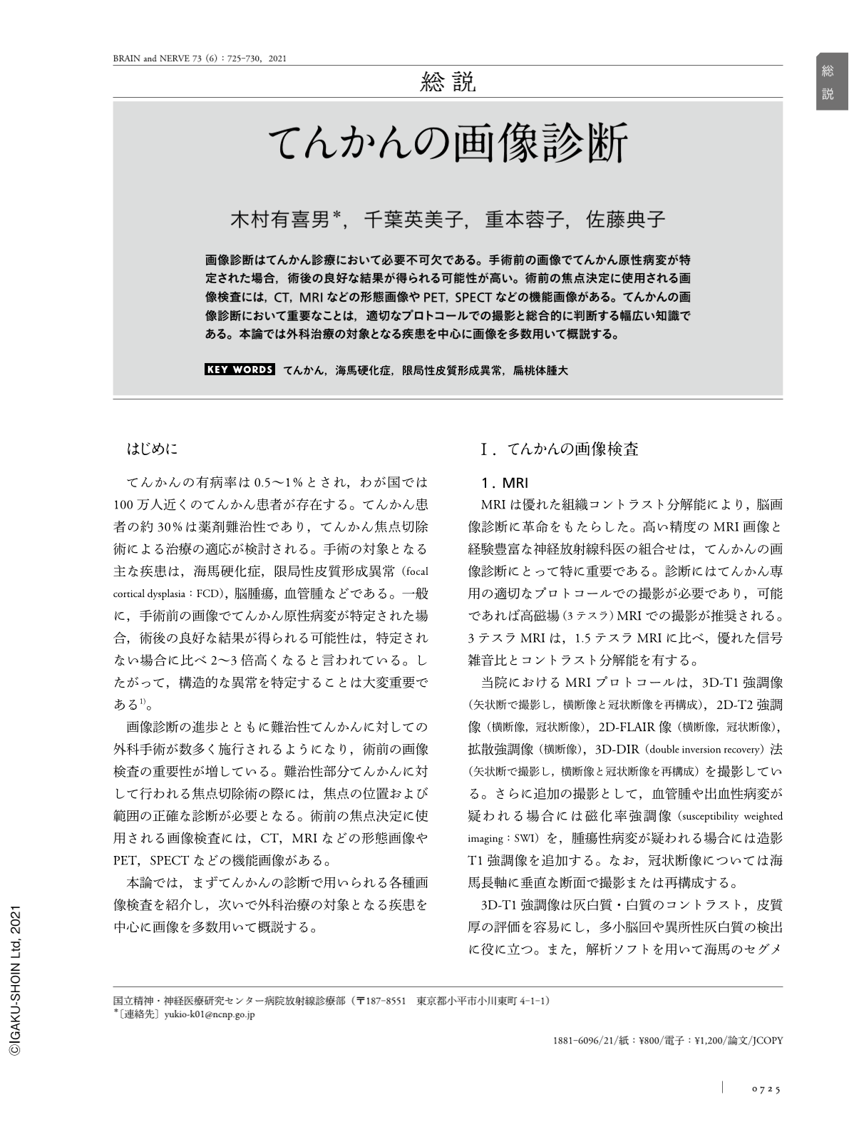
画像診断はてんかん診療において必要不可欠である。手術前の画像でてんかん原性病変が特定された場合,術後の良好な結果が得られる可能性が高い。術前の焦点決定に使用される画像検査には,CT,MRIなどの形態画像やPET,SPECTなどの機能画像がある。てんかんの画像診断において重要なことは,適切なプロトコールでの撮影と総合的に判断する幅広い知識である。本論では外科治療の対象となる疾患を中心に画像を多数用いて概説する。
Abstract
Neuroimaging is the most important tool to treat epileptic foci. Preoperative detection of underlying structural lesions increases the likelihood of the successful surgical treatment of drug-resistant epilepsy. Imaging studies used for preoperative focal determination include morphological imaging modalities, such as computed tomography and magnetic resonance imaging, and functional ones, such as positron emission tomography and single photon-emission computed tomography. The keys to diagnostic imaging in epilepsy are appropriate protocols and extensive knowledge to make comprehensive decisions. This article comprehensively overviews neuroimaging techniques used in epilepsy.
Copyright © 2021, Igaku-Shoin Ltd. All rights reserved.