Japanese
English
- 有料閲覧
PPV購入案内
1,320円 (1,200円+税10%) こちらは、359ページから362ページの文献です。購入には医書.jp本会員登録が必要です。
会員登録完了後に改めて上記「購入サイトへ」を選択してください。
または「購入サイトへ」選択後に「購入ログイン」、「新規会員登録」の順に進んでください。
会員登録について詳しくはをご確認ください。
- Abstract 文献概要
- 1ページ目 Look Inside
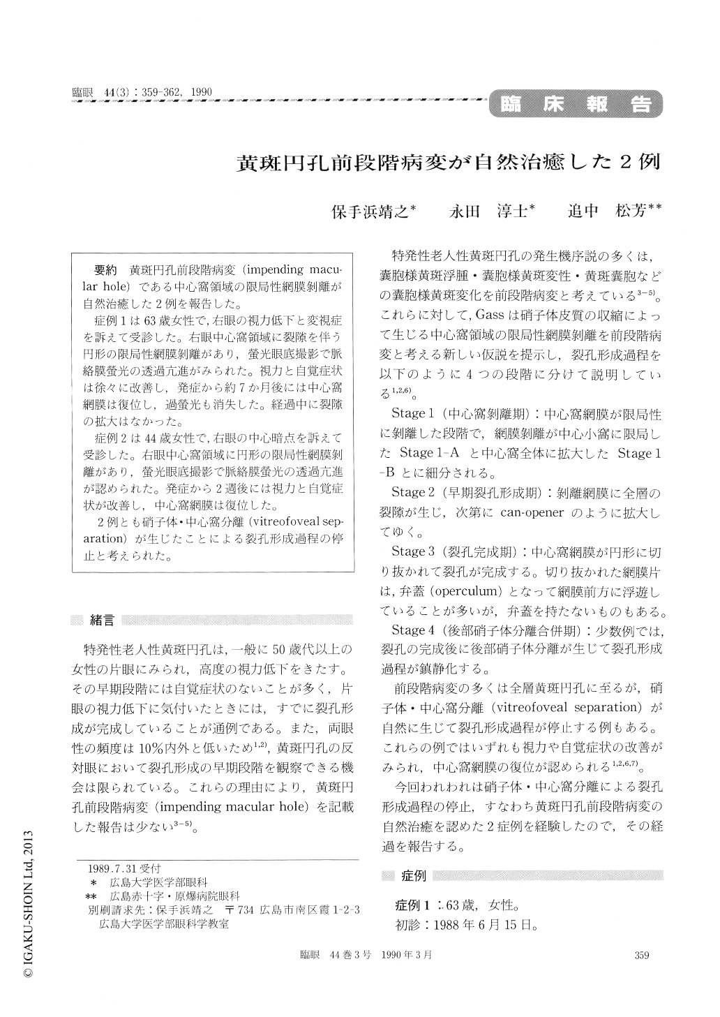
黄斑円孔前段階病変(impending macu-lar hole)である中心窩領域の限局性網膜剥離が自然治癒した2例を報告した。
症例1は63歳女性で,右眼の視力低下と変視症を訴えて受診した。右眼中心窩領域に裂隙を伴う円形の限局性網膜剥離があり,螢光眼底撮影で脈絡膜螢光の透過亢進がみられた。視力と自覚症状は徐々に改善し,発症から約7か月後には中心窩網膜は復位し,過螢光も消失した。経過中に裂隙の拡大はなかった。
症例2は44歳女性で,右眼の中心暗点を訴えて受診した。右眼中心窩領域に円形の限局性網膜剥離があり,螢光眼底撮影で脈絡膜螢光の透過亢進が認められた。発症から2週後には視力と自覚症状が改善し,中心窩網膜は復位した。
2例とも硝子体・中心窩分離(vitreofoveal sep-aration)が生じたことによる裂孔形成過程の停止と考えられた。
Two females, aged 63 and 44 years each, showed spontaneous resolution of impending macular hole manifesting focal retinal detachment in the foveal area. We observed, as common features in both, a round lesion corresponding to focal retinal detach-ment in the fovea and increased transmission of choroidal fluorescence in this lesion. Only one eye was affected in both cases. The visual acuity in the involved eye measured 0.3 and 1.0 respectively. Spontaneous improvement resulted after 7 months in one case and after 2 weeks in the other, resulting in disappearance of subjective symptoms and retinal reattachment at the fovea. The visual acuity improved to 1.0 and 1.5 respectively. Spontaneous vitreofoveal separation seemed to be the cause of observed arrest and improvement of impending macular hole in both cases.
Copyright © 1990, Igaku-Shoin Ltd. All rights reserved.