Japanese
English
- 有料閲覧
PPV購入案内
1,320円 (1,200円+税10%) こちらは、78ページから80ページの文献です。購入には医書.jp本会員登録が必要です。
会員登録完了後に改めて上記「購入サイトへ」を選択してください。
または「購入サイトへ」選択後に「購入ログイン」、「新規会員登録」の順に進んでください。
会員登録について詳しくはをご確認ください。
- Abstract 文献概要
- 1ページ目 Look Inside
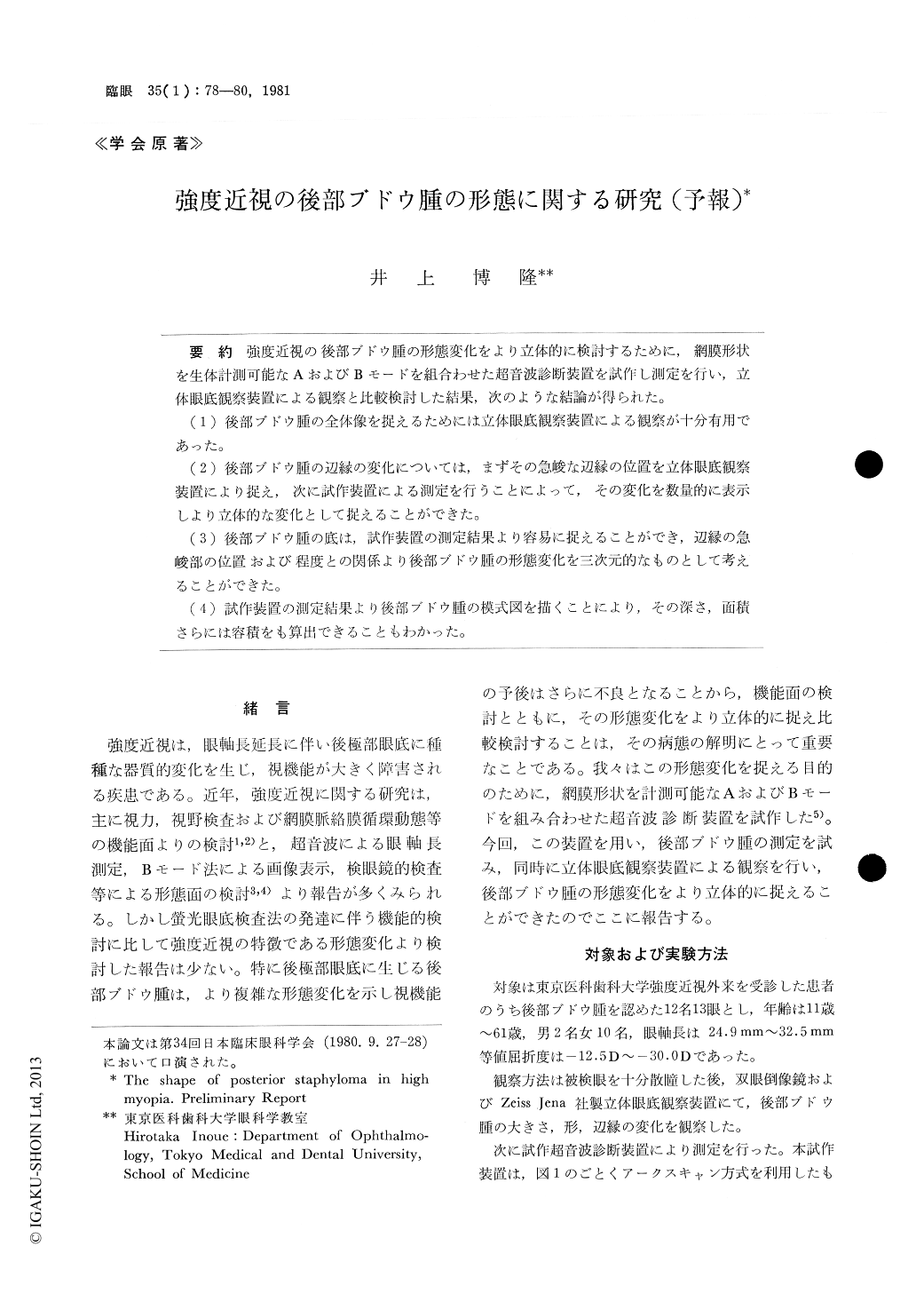
強度近視の後部ブドウ腫の形態変化をより立体的に検討するために,網膜形状を生体計測可能なAおよびBモードを組合わせた超音波診断装置を試作し測定を行い,立体眼底観察装置による観察と比較検討した結果,次のような結論が得られた。
(1)後部ブドウ腫の全体像を捉えるためには立体眼底観察装置による観察が十分有用であった。
(2)後部ブドウ腫の辺縁の変化については,まずその急峻な辺縁の位置を立体眼底観察装置により捉え,次に試作装置による測定を行うことによって,その変化を数量的に表示しより立体的な変化として捉えることができた。
(3)後部ブドウ腫の底は,試作装置の測定結果より容易に捉えることができ,辺縁の急峻部の位置および程度との関係より後部ブドウ腫の形態変化を三次元的なものとして考えることができた。
(4)試作装置の測定結果より後部ブドウ腫の模式図を描くことにより,その深さ,面積さらには容積をも算出できることもわかった。
We constructed an ultrasonographic apparatus using an arc-scan method. This enabled a digital measurement of the distance from the apex of the cornea to the retinal surface for each degree of the arc scan. This method was applied to 13 eyes with posterior staphyloma due to high myopia. A fairly accurate and objective measurement of the staphy-loma could be performed. The depth of staphy-loma was inversely correlated with the best cor-rected visual acuity.
Copyright © 1981, Igaku-Shoin Ltd. All rights reserved.