Japanese
English
- 有料閲覧
PPV購入案内
1,320円 (1,200円+税10%) こちらは、983ページから987ページの文献です。購入には医書.jp本会員登録が必要です。
会員登録完了後に改めて上記「購入サイトへ」を選択してください。
または「購入サイトへ」選択後に「購入ログイン」、「新規会員登録」の順に進んでください。
会員登録について詳しくはをご確認ください。
- Abstract 文献概要
- 1ページ目 Look Inside
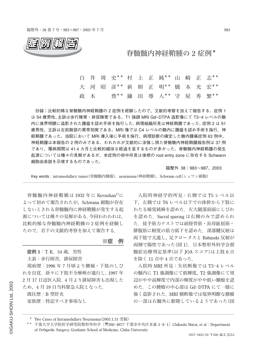
抄録:比較的稀な脊髄髄内神経鞘腫の2症例を経験したので,文献的考察を加えて報告する.症例1は54歳男性,主訴は歩行障害・排尿障害である.T1強調MRI Gd-DTPA造影像にてT3-4レベルの髄内に境界明瞭に造影された腫瘤を認め手術を施行した.病理組織所見は神経鞘腫であった.症例2は51歳男性,主訴は左前腕部の異常知覚である.MRI像ではC4レベルの髄内に腫瘤を認め手術を施行,神経鞘腫であった.当院においてMRI導入後に手術を施行,病理診断の確定した髄内腫瘍症例63例中,神経鞘腫は本報告の2例のみである.われわれが文献的に渉猟し得た脊髄髄内神経鞘腫報告例は37例であり,罹病期間は41.4カ月と比較的緩徐な経過を呈するものが多かった.脊髄髄内神経鞘腫の発生起源については種々の見解があるが,本症例の術中所見は後根のroot entry zoneに存在するSchwann細胞由来説を示唆するものであった.
Intramedullary neurinoma of the spinal cord has been rarely reported. We report two additional cases of intramedullary neurinoma of the spinal cord and review the literature. Case 1;A 54-year-old man, who had gait disturbance and dysuresia. Magnetic resonance imaging (MRI) with gadolinium revealed Intramedullary spinal tumor at the third and fourth thoracic segments. We operated on the patient. Histology of the tumor was neurinoma showing spindle cells arranged in palisading fashion. Case 2;A 51-year-old man, who had dysesthesia of the left forearm. MRI with gadolinium revealed intramedullary spinal tumor at the fourth cervical segment. Histological examination of the tumor revealed spindle cells arranged in palisading fashion. The histology of both tumors was the same. The standard textbook on tumors of the central nervous system do not even mention the existence of Schwann cell tumor within the parenchyma of the spinal cord. We suggests these tumors originate and grow centripetally from the dorsal nerve root entry zone, taking into account the tumor locations at surgery.
Copyright © 2003, Igaku-Shoin Ltd. All rights reserved.