Japanese
English
- 有料閲覧
PPV購入案内
1,320円 (1,200円+税10%) こちらは、1163ページから1168ページの文献です。購入には医書.jp本会員登録が必要です。
会員登録完了後に改めて上記「購入サイトへ」を選択してください。
または「購入サイトへ」選択後に「購入ログイン」、「新規会員登録」の順に進んでください。
会員登録について詳しくはをご確認ください。
- Abstract 文献概要
- 1ページ目 Look Inside
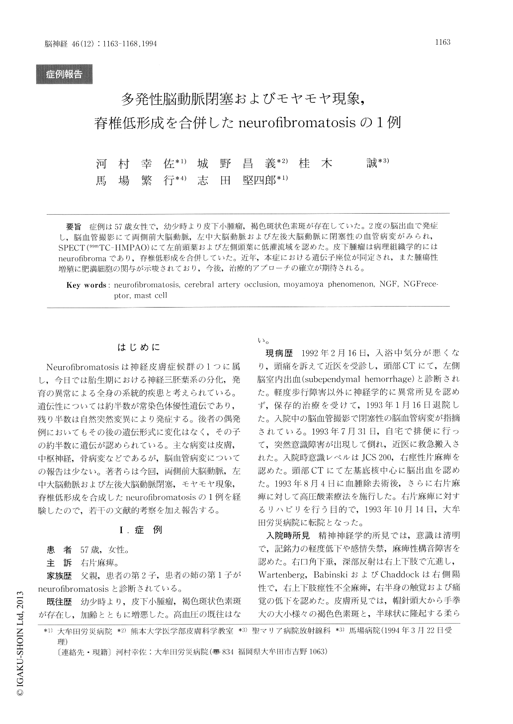
症例は57歳女性で,幼少時より皮下小腫瘤,褐色斑状色素斑が存在していた。2度の脳出血で発症し,脳血管撮影にて両側前大脳動脈,左中大脳動脈および左後大脳動脈に閉塞性の血管病変がみられ,SPECT(99mTC-HMPAO)にて左前頭葉および左側頭葉に低灌流域を認めた。皮下腫瘤は病理組織学的にはneurofibromaであり,脊椎低形成を合併していた。近年,本症における遺伝子座位が同定され,また腫瘍性増殖に肥満細胞の関与が示唆されており,今後,治療的アプローチの確立が期待される。
Cases of cerebrovascular occlusive lesion with neurofibromatosis have rarely been reported. We report here, the case of a 57-year-old woman who twice had sudden onset of brain hemorrhage. She had a family history of neurofibromatosis. Her elder daughter and her niece had a diagnosis of neurofibromatosis and examination showed cafe au lait spots and neurofibroma over the body, ac-companied with scoliosis. CT scans revealed sube-pendymal hemorrhage at the first onset of cerebral bleeding and secondly showed a high-density mass at the left basal ganglia.
Copyright © 1994, Igaku-Shoin Ltd. All rights reserved.