Japanese
English
- 有料閲覧
PPV購入案内
1,320円 (1,200円+税10%) こちらは、1217ページから1223ページの文献です。購入には医書.jp本会員登録が必要です。
会員登録完了後に改めて上記「購入サイトへ」を選択してください。
または「購入サイトへ」選択後に「購入ログイン」、「新規会員登録」の順に進んでください。
会員登録について詳しくはをご確認ください。
- Abstract 文献概要
- 1ページ目 Look Inside
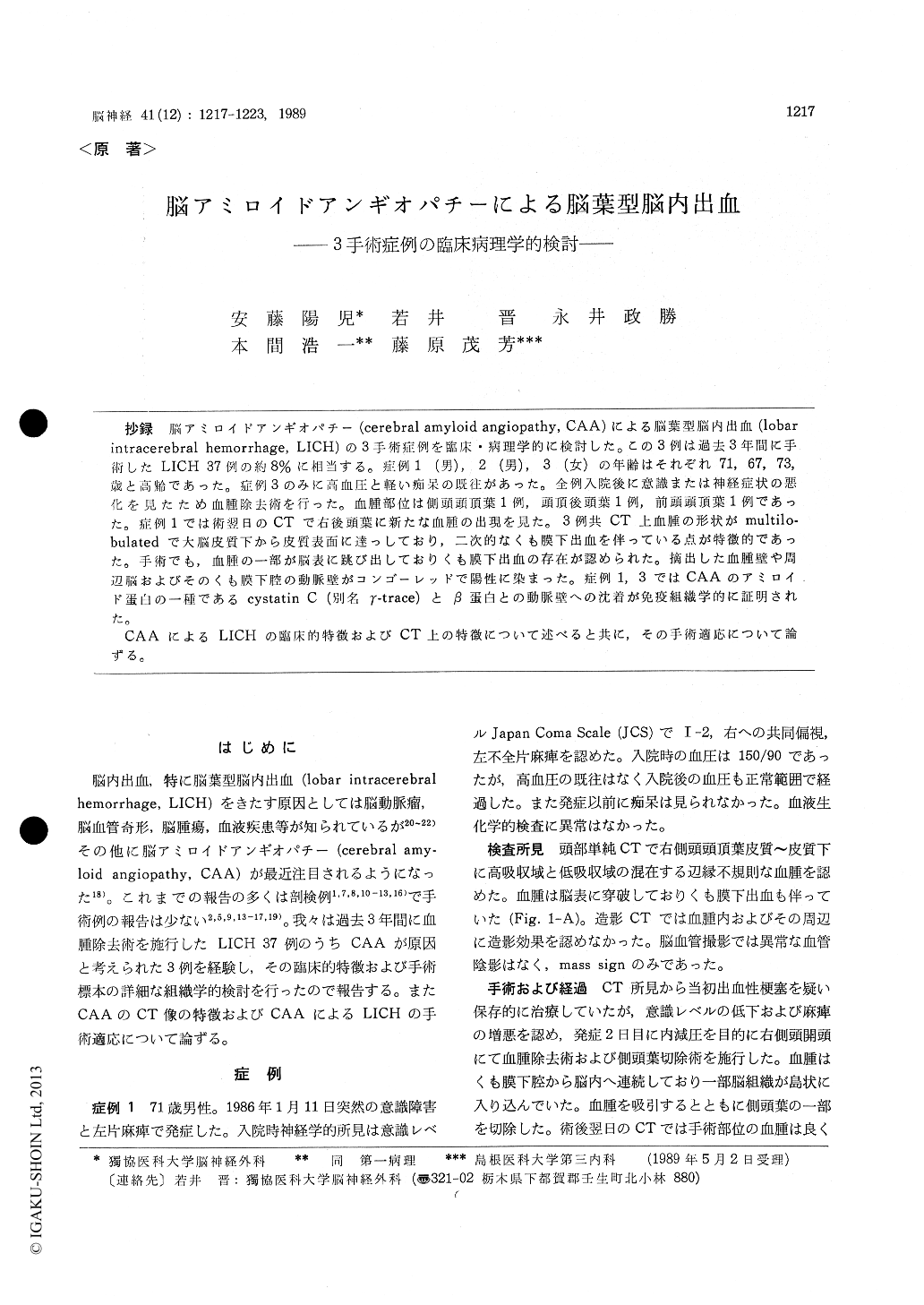
抄録 脳アミロイドアンギオパチー(cerebral amyloid angiopathy, CAA)による脳葉型脳内出血(10bar intracerebral hemorrhage, LICH)の3手術症例を臨床・病理学的に検討した。この3例は過去3年間に手術したLICH 37例の約8%に相当する。症例1(男),2(男),3(女)の年齢はそれぞれ71,67,73,歳と高齢であった。症例3のみに高血圧と軽い痴呆の既往があった。全例入院後に意識または神経症状の悪化を見たため血腫除去術を行った。血腫部位は側頭頭頂葉1例,頭頂後頭葉1例,前頭頭頂葉1例であった。症例1では術翌日のCTで右後頭葉に新たな血腫の出現を見た。3例共CT上血腫の形状がmultilo-bulatedで大脳皮質下から皮質表面に達っしており,二次的なくも膜下出血を伴っている点が特徴的であった。手術でも,血腫の一部が脳表に跳び出しておりくも膜下出血の存在が認められた。摘出した血腫壁や周辺脳およびそのくも膜下腔の動脈壁がコンゴーレッドで陽性に染まった。症例1,3ではCAAのアミロイド蛋白の一種であるcystatin C (別名γ-trace)とβ蛋白との動脈壁への沈着が免疫組織学的に証明された。
CAAによるLICHの臨床的特徴およびCT上の特徴について述べると共に,その手術適応について論ずる。
Three operated cases of lobar intracerebral hemorrhage (LICH) related to cerebral amyloid angiopathy (CAA) were studied clinicopatholo-gically. They constituted about 8% of all LICH cases (n=37) operated upon in our institute (DUSM) during the past 3 years. Case 1, 2 and 3 aged 71, 67 and 73 years, respectively. There were 2 males (Cases 1 & 2) and 1 female (Case 3). Only one case (Case 3) had both hypertension and dementia before hemorrhage. In all 3 cases, neurologic symptoms deteriorated after admission. The hematoma involved the right temporo-parietal in 1 (Case 1), the right parieto-occipital in 1 (Case 2) and the left fronto-parietal region in 1 (Case 3). Case 1 developed a new hematoma in the right occipital lobe on the day following surgery. On CT, the hematoma was multilobulated in shape and located very superficially extending to the subarachnoid space in all cases. There was no abnormal enhancement in and around the hematoma upon contrast infusion. Angiography showed only an avascular mass sign in case. At surgery, the hematoma was extruded onto the cortical surface in all cases. The surgical outcome was good in 2 (Cases 1 & 2) and fair in 1 (Case 3).
Removed hematomas, solid nodular tissues and adjacent brain tissues were examined histological-ly using hematoxylin and eosin, Azan-Mallory, elastica van Gieson, silver and Congo red stains.Arteries in the hematoma wall, the subarachnoid space and the adjacent brain parenchyma were intensely stained with Congo red and showed birefringence on polarized light. In the periphery of the hematoma removed from Case 3, there was a microaneurysm of which wall was also stained with Congo red. In 2 cases (Cases I & 3), deposition of cystatin C (gamma-trace) and beta protein, two of the CAA related amyloid proteins, was investigated immunohistochemically using the ABC method. Dense reaction products for bothamyloid proteins were observed in the arterial wall in both cases.
When the CAA is strongly suspected as a cause of a given LICH based upon the clinical and CT features, surgical evacuation of the hematoma should not be considered as a treatment option unless the neurological symptoms deteriorate. The operative indication for LICH is discussed in this regard.
Copyright © 1989, Igaku-Shoin Ltd. All rights reserved.