Japanese
English
- 有料閲覧
PPV購入案内
1,320円 (1,200円+税10%) こちらは、259ページから263ページの文献です。購入には医書.jp本会員登録が必要です。
会員登録完了後に改めて上記「購入サイトへ」を選択してください。
または「購入サイトへ」選択後に「購入ログイン」、「新規会員登録」の順に進んでください。
会員登録について詳しくはをご確認ください。
- Abstract 文献概要
- 1ページ目 Look Inside
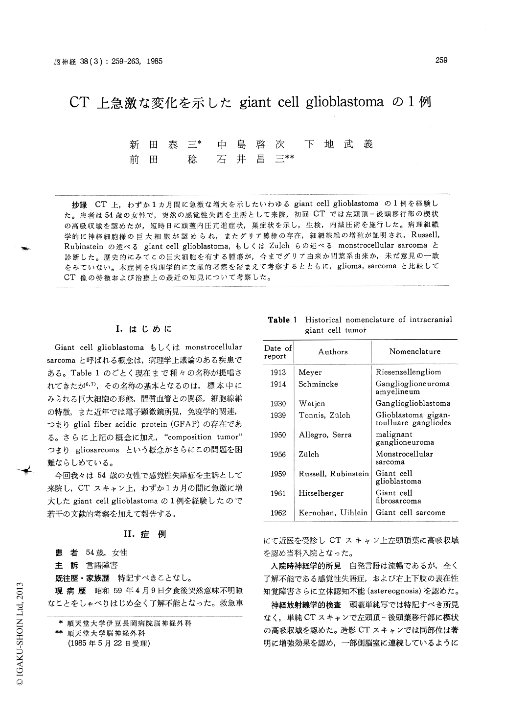
抄録 CT上,わずか1カ月間に急激な増大を示したいわゆるgiant cell glioblastomaの1例を経験した。患者は54歳の女性で,突然の感覚性失語を主訴として来院,初回CTでは左頭頂—後頭移行部の楔状の高吸収域を認めたが,短時日に頭蓋内圧亢進症状,巣症状を示し,生検,内減圧術を施行した。病理組織学的に神経細胞様の巨大細胞が認められ,またグリア線維の存在,細網線維の増殖が証明され,Russell,Rubinsteinの述べるgiant cell glioblastoma,もしくはZülchらの述べるmonstrocellular sarcomaと診断した。歴史的にみてこの巨大細胞を有する腫瘍が,今までグリア由来か間葉系由来か,未だ意見の一致をみていない。本症例を病理学的に文献的考察を踏まえて考察するとともに,glioma, sarcomaと比較してCT像の特徴および治療上の最近の知見について考察した。
The pathological entity of intracranial giant cell tumor remains controversial, which has been considered a glioma (giant cell glioblastoma) by some and a sarcoma (monstrocellular sarcoma) by others. The authors present a case in which CT scan shows a wedge-shaped high density area at the left parietal lobe. This tumor expands explo-sively only a month on serial CT scan. The mic-roscopic speciemen demonstrates the bizarre ir-regular giant cells with large vesicular nuclei, and glial proliferation and abundant reticulin fibers. Then we reviewed and discussed with emphasis on the historical aspect of giant cell tumor and also radiologic, therapeutic problem.
Copyright © 1986, Igaku-Shoin Ltd. All rights reserved.