Japanese
English
- 有料閲覧
PPV購入案内
1,320円 (1,200円+税10%) こちらは、1145ページから1154ページの文献です。購入には医書.jp本会員登録が必要です。
会員登録完了後に改めて上記「購入サイトへ」を選択してください。
または「購入サイトへ」選択後に「購入ログイン」、「新規会員登録」の順に進んでください。
会員登録について詳しくはをご確認ください。
- Abstract 文献概要
- 1ページ目 Look Inside
I.はじめに
大脳の皮質あるいは皮質直下に小出血巣をみる例は,従来より剖検により偶然発見され12,13),高血圧症や脳動脈硬化症と関連するものとか,あるいはsmall angio—matous malformation (SAM)や紡錘形の脳動脈瘤の破綻によるものとされてきた2,3,12,22)。すなわち,このような例は剖検により発見されるのみで,脳血管写,脳波あるいは,放射性同位元素などによる従来の臨床的検査法で,臨床的に診断することはできなかつたものといえよう。
さて,われわれは外来診察中の患者で,late onsetepilepsyとされた症例の中に,CT (computed tomo—graphy)によりおそらくは皮質あるいは皮質直下に小出血巣をもつ,いわゆる皮質近傍小出血の8例を経験し,そのうち3例は開頭手術によりこれを確認した。このような例はCTによりはじめて臨床診断が可能となつたものと考えられる。
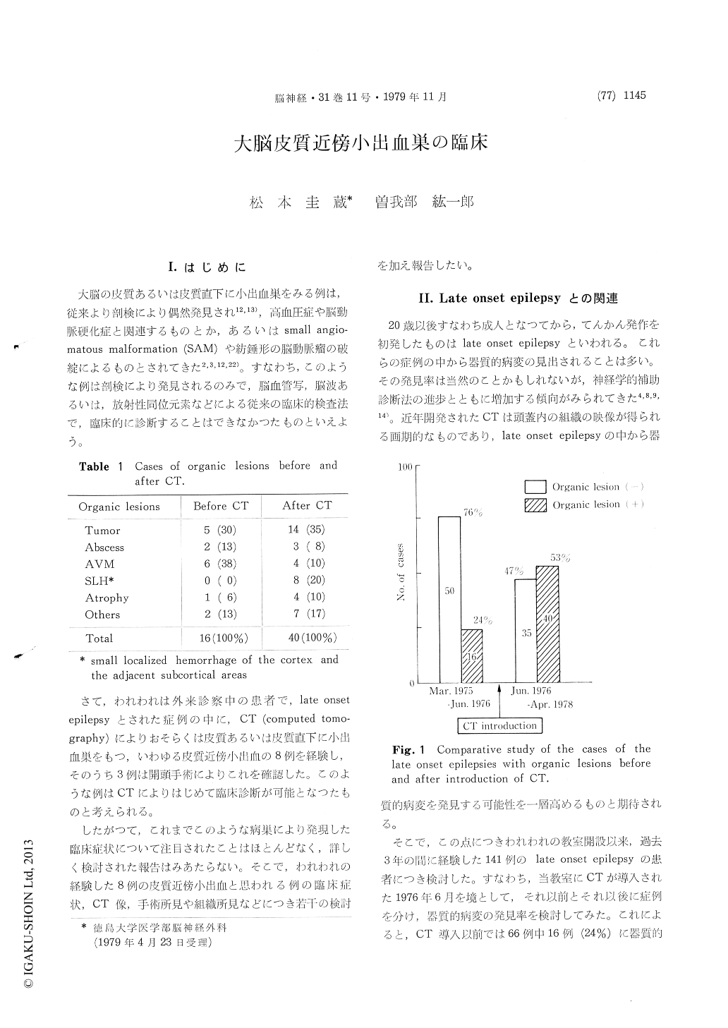
A consecutive series of 141 cases of late onset epilepsies has been treated in our service for last 3 years. Utilization of computed tomography (CT) for the examination of those cases was started from June 1976. Organic lesions were detected 16 cases (24%) out of a series of 66 cases without CT examination and 40 cases (53%) out of a series of 75 cases in which the CT examinations were applied.
It was very interesting that there were 8 cases of a small localized hemorrhages in the cerebral cortex and the adjacent subcortical area, which was noticed only by CT studies in the latter series. Ages of these cases were ranged from 32 to 64 years old with 5 cases of male and 3 cases of female. Chief complaint was episode of convulsive attack in all cases. Initiations of the disease were also partial seizures in 2 cases and generalized seizures in 5 cases. One other case had some transient articulation disturbances as an initial symptom. 4 cases had attacks in the early morning when asleep out of 5 case who had generalizedseizure as an initial symptom. History of hyper-tension was noted in 3 cases of 6 decades. The other 5 cases had no significant history of illness. There were no neurological deficits in the routine examination at the time of intervals of their attacks in all cases. EEG examination of these cases revealed abnormalities of focal slow or sporadic slow wave in the majority of the cases. However, there was no case whose localization of EEG focus corresponded with the location of the lesions visualized by CT.
CT appearances of the lesions were quite char-acteristic. A small round spotted or patched high density area with irregular margin were noted in the cortex or the adjacent subcortical areas by plain CT study. There was neither surrounding low density area nor enhancement effect by intra-venous injection of the contrast media. Moreover, the high density area maintained similar shape for over 3 months long by follow-up CT studies in some cases.
3 cases under-went craniotomies and their lesions were totally removed. The histological examinations revealed old hemorrhagic glia cicatrices in all cases, and suggested us that the causes of lesions were a small hemorrhagic infarction in two cases and a small hemorrhages possibly from small angiomatous malformations, of which Morgolis cited, in the other case.
It has been already known that there are a small localized cortical hemorrhages arising from a small angiomatous malformation or a cryptic fusiform aneurysm of the cortex, which had been detected incidentally at the postmortem examination. However, it seemed to have been paid no attention to the clinical features of these small hemorrhages.
Now, it has been stressed that these small hemor-rhagic lesions may provoke an epileptic seizure as a cardinal clinical symptom and CT examination will elucidate these cases clinically among the cases of late onset epilepsy, although exsistence of asymptomatic cases may not be ruled out.
Copyright © 1979, Igaku-Shoin Ltd. All rights reserved.