Japanese
English
- 有料閲覧
PPV購入案内
1,320円 (1,200円+税10%) こちらは、429ページから433ページの文献です。購入には医書.jp本会員登録が必要です。
会員登録完了後に改めて上記「購入サイトへ」を選択してください。
または「購入サイトへ」選択後に「購入ログイン」、「新規会員登録」の順に進んでください。
会員登録について詳しくはをご確認ください。
- Abstract 文献概要
- 1ページ目 Look Inside
- 参考文献 Reference
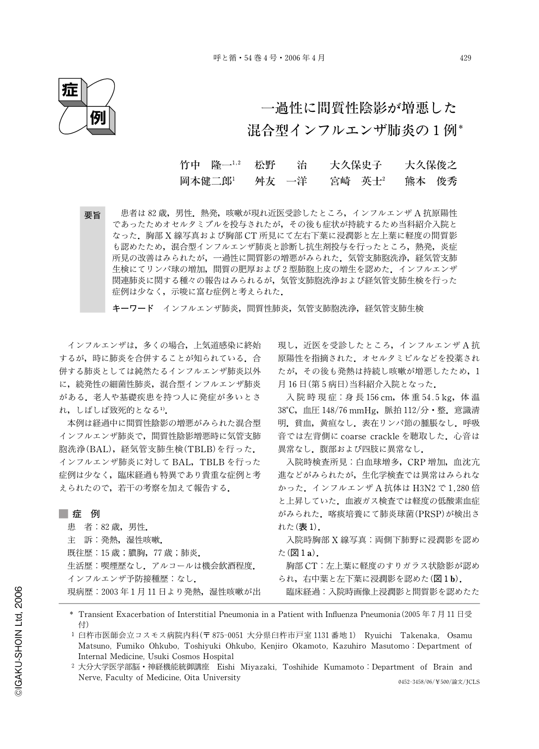
患者は82歳,男性.熱発,咳嗽が現れ近医受診したところ,インフルエンザA抗原陽性であったためオセルタミブルを投与されたが,その後も症状が持続するため当科紹介入院となった.胸部X線写真および胸部CT所見にて左右下葉に浸潤影と左上葉に軽度の間質影も認めたため,混合型インフルエンザ肺炎と診断し抗生剤投与を行ったところ,熱発,炎症所見の改善はみられたが,一過性に間質影の増悪がみられた.気管支肺胞洗浄,経気管支肺生検にてリンパ球の増加,間質の肥厚および2型肺胞上皮の増生を認めた.インフルエンザ関連肺炎に関する種々の報告はみられるが,気管支肺胞洗浄および経気管支肺生検を行った症例は少なく,示唆に富む症例と考えられた.
A 82-year-old man with influenza infection was admitted to our hospital because of fever and cough. Chest radiograph and CT showed infiltrates in both lung and ground glass opacity in the left upper lobe. Five days after admission, chest CT showed deterioration of ground glass opacity. Bronchoalveolar lavage analysis showed increase in the number of lymphocytes and decrease of CD4+ to CD8+ rate. Transbronchial lung biopsy showed that the alveolar septa were thickend by a combination of inflammation, fibroblast proliferation, and alveolar pneumocyte hyperplasia. The interstitial pneumonia gradually improved without treatment. This was a rare case of influenza pneumonia which showed transient deterioration of interstitial change.
Copyright © 2006, Igaku-Shoin Ltd. All rights reserved.