Japanese
English
- 有料閲覧
PPV購入案内
1,320円 (1,200円+税10%) こちらは、888ページから895ページの文献です。購入には医書.jp本会員登録が必要です。
会員登録完了後に改めて上記「購入サイトへ」を選択してください。
または「購入サイトへ」選択後に「購入ログイン」、「新規会員登録」の順に進んでください。
会員登録について詳しくはをご確認ください。
- Abstract 文献概要
- 1ページ目 Look Inside
- 参考文献 Reference
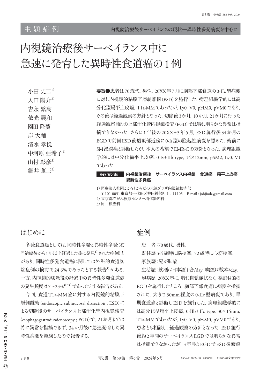
要旨●患者は70歳代,男性.20XX年7月に胸部下部食道の0-IIc型病変に対し内視鏡的粘膜下層剝離術(ESD)を施行した.病理組織学的には高分化型扁平上皮癌,T1a-MMであったが,Ly0,V0,pHM0,pVM0であり,その後は経過観察の方針となった.切除後3か月,10か月,21か月に行った経過観察目的の上部消化管内視鏡検査(EGD)では特に明らかな異常は指摘できなかった.さらに1年後の20XX+3年5月,ESD施行後34か月のEGDで前回ESD後瘢痕部近傍に0-Is型の隆起性病変を認めた.術前にSM浸潤癌と診断したが,本人の希望でEMR-Cの方針となった.病理組織学的には中分化扁平上皮癌,0-Is+IIb type,14×12mm,pSM2,Ly0,V1であった.
In July 20XX, a 70-year-old male underwent ESD(endoscopic submucosal dissection)for type 0-IIc squamous cell carcinoma in the lower thoracic esophagus. Histopathological findings revealed well-differentiated squamous cell carcinoma, classified as T1a-MM, Ly0, V0, HM0, and VM0, leading to a decision for observational follow-up. EGD(esophagogastroduodenoscopy)conducted 3 months, 10 months, and 21 months after resection showed no significant abnormalities. Furthermore, 1 year later, in May 20XX+3, 34 months after treatment, a 0-Is type lesion was found near the scar following the previous ESD. Although the preoperative diagnosis was SM invasive cancer, EMR-C was decided based on the patient's wish. Histopathological findings showed a moderately differentiated squamous cell carcinoma, classified as 0-Is + IIb type, 14×12mm, pT1b-SM2, Ly0, V1.
Copyright © 2024, Igaku-Shoin Ltd. All rights reserved.