Japanese
English
- 有料閲覧
PPV購入案内
1,320円 (1,200円+税10%) こちらは、1727ページから1733ページの文献です。購入には医書.jp本会員登録が必要です。
会員登録完了後に改めて上記「購入サイトへ」を選択してください。
または「購入サイトへ」選択後に「購入ログイン」、「新規会員登録」の順に進んでください。
会員登録について詳しくはをご確認ください。
- Abstract 文献概要
- 1ページ目 Look Inside
- 参考文献 Reference
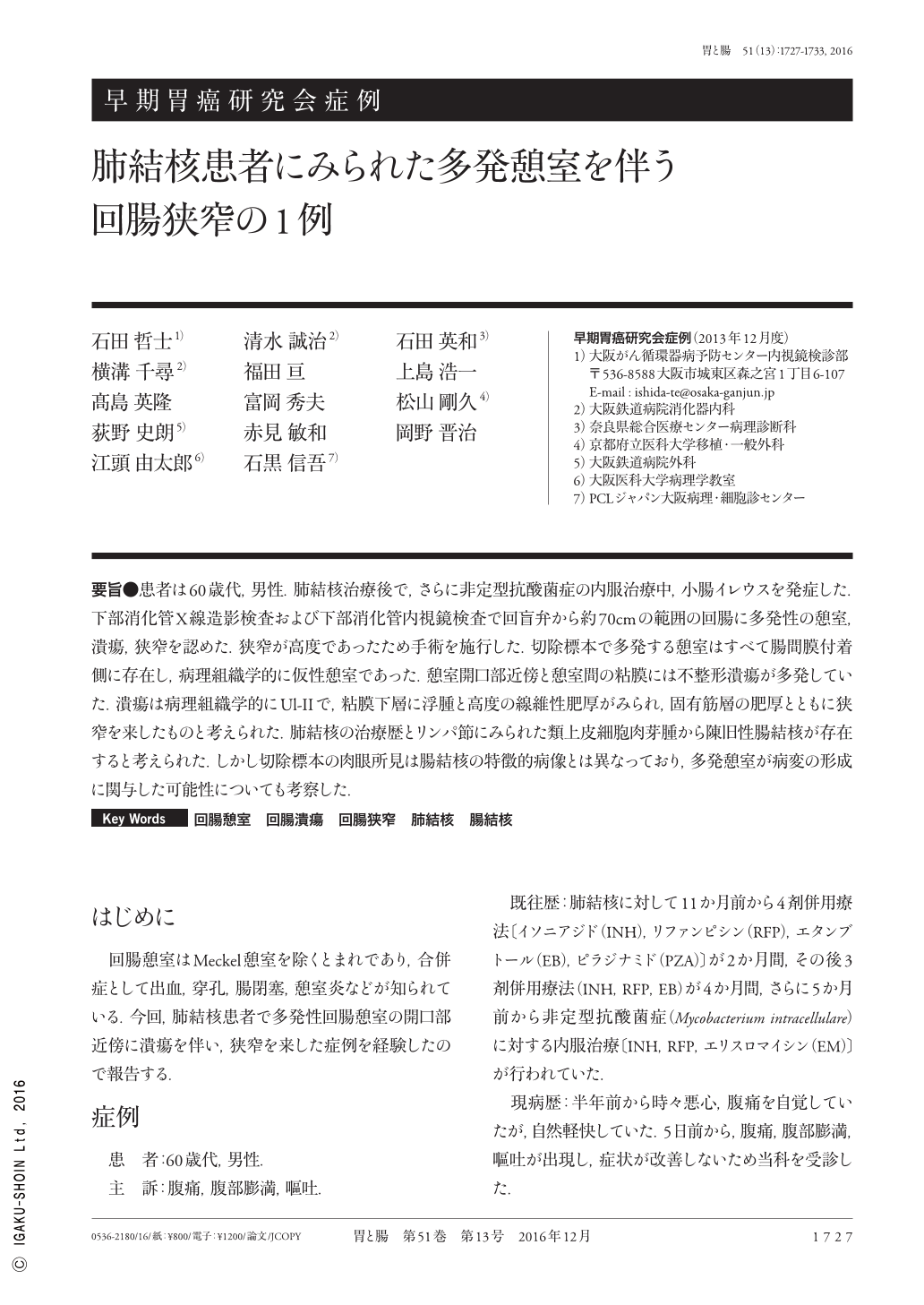
要旨●患者は60歳代,男性.肺結核治療後で,さらに非定型抗酸菌症の内服治療中,小腸イレウスを発症した.下部消化管X線造影検査および下部消化管内視鏡検査で回盲弁から約70cmの範囲の回腸に多発性の憩室,潰瘍,狭窄を認めた.狭窄が高度であったため手術を施行した.切除標本で多発する憩室はすべて腸間膜付着側に存在し,病理組織学的に仮性憩室であった.憩室開口部近傍と憩室間の粘膜には不整形潰瘍が多発していた.潰瘍は病理組織学的にUl-IIで,粘膜下層に浮腫と高度の線維性肥厚がみられ,固有筋層の肥厚とともに狭窄を来したものと考えられた.肺結核の治療歴とリンパ節にみられた類上皮細胞肉芽腫から陳旧性腸結核が存在すると考えられた.しかし切除標本の肉眼所見は腸結核の特徴的病像とは異なっており,多発憩室が病変の形成に関与した可能性についても考察した.
A 68-year-old man, who had been treated for atypical mycobacterial infection following termination of treatment for pulmonary tuberculosis, presented with small intestinal ileus. Radiographic and endoscopic examinations revealed multiple diverticuli, ulcers, and stenoses in the ileum, located within 70cm of the ileocecal valve. Surgery was performed because of severe stenoses. The resected specimen showed multiple pseudodiverticuli that were exclusively lined on the mesenteric border Irregularly shaped ulcers were found to be distributed around and between the diverticuli. Ulcers were approximately UL-II in depth and were accompanied by edema and dense submucosal fibrosis. Stenoses were considered to be caused by wall thickening due to submucosal fibrosis and hypertrophy of the proper muscle layer. The presence of small, noncaseating epithelioid granulomas in the lymph nodes was suggestive of healed intestinal tuberculosis. However, the macroscopic appearances of ileal lesions were different from those of intestinal tuberculosis. Instead, ileal diverticuli may have been related to the formation of ulcers and stenoses in this patient.
Copyright © 2016, Igaku-Shoin Ltd. All rights reserved.