Japanese
English
- 有料閲覧
PPV購入案内
1,320円 (1,200円+税10%) こちらは、1255ページから1266ページの文献です。購入には医書.jp本会員登録が必要です。
会員登録完了後に改めて上記「購入サイトへ」を選択してください。
または「購入サイトへ」選択後に「購入ログイン」、「新規会員登録」の順に進んでください。
会員登録について詳しくはをご確認ください。
- Abstract 文献概要
- 1ページ目 Look Inside
- 参考文献 Reference
- サイト内被引用 Cited by
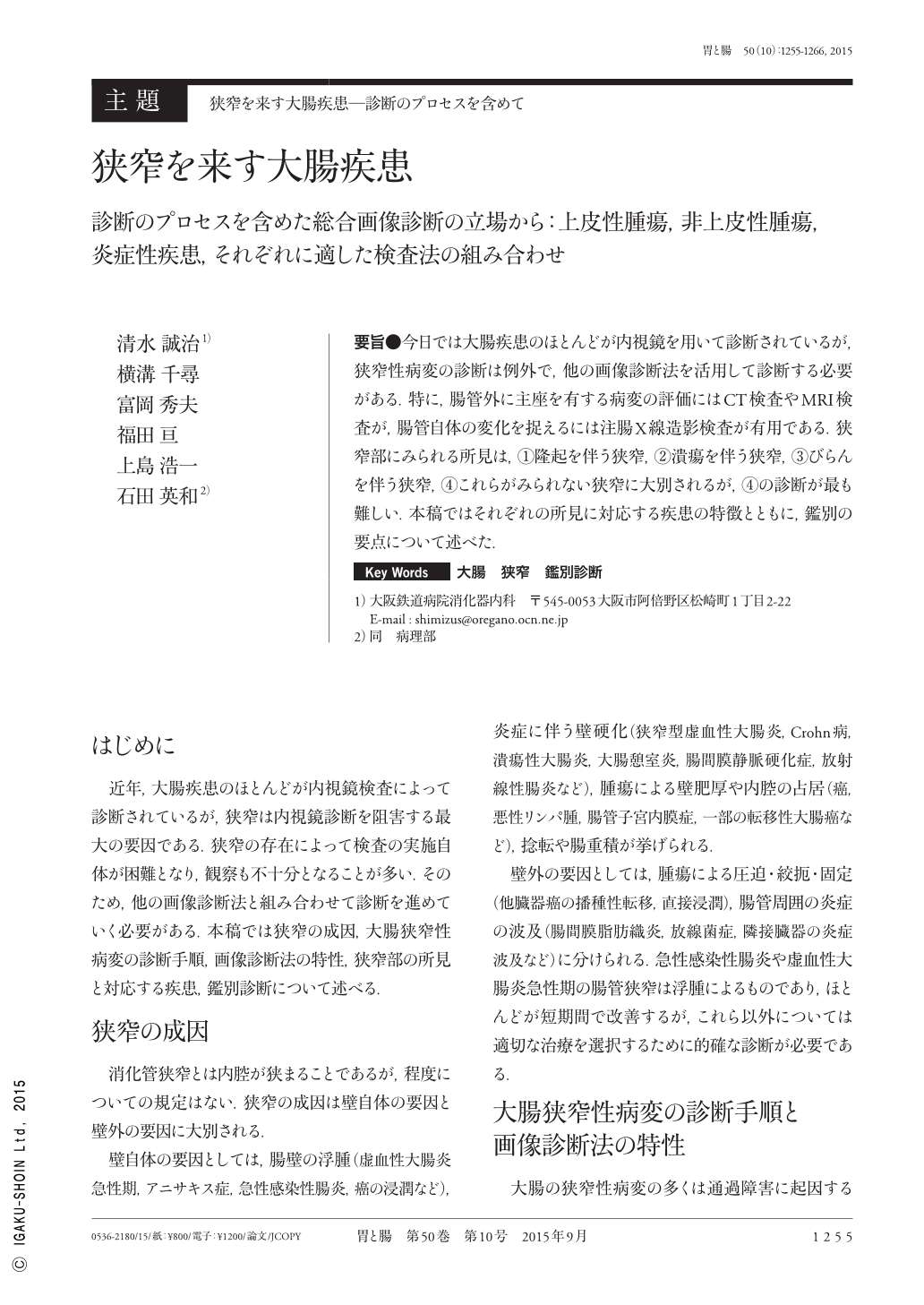
要旨●今日では大腸疾患のほとんどが内視鏡を用いて診断されているが,狭窄性病変の診断は例外で,他の画像診断法を活用して診断する必要がある.特に,腸管外に主座を有する病変の評価にはCT検査やMRI検査が,腸管自体の変化を捉えるには注腸X線造影検査が有用である.狭窄部にみられる所見は,(1)隆起を伴う狭窄,(2)潰瘍を伴う狭窄,(3)びらんを伴う狭窄,(4)これらがみられない狭窄に大別されるが,(4)の診断が最も難しい.本稿ではそれぞれの所見に対応する疾患の特徴とともに,鑑別の要点について述べた.
At present, most colorectal diseases are diagnosed by colonoscopy ; however, the diagnosis of diseases causing stenosis often becomes possible only by employment of other imaging modalities such as barium enema, CT, MRI, and ultrasonography. Of these, CT is useful for evaluation of extramural changes and barium enema for evaluation of mural ones. The findings related to stenotic lesions are classified into four categories : 1)stenoses with elevated lesions, 2)stenoses with ulcers, 3)stenoses with erosions, and 4)stenoses without such changes. Of them, the last category is the most difficult to diagnose. In this article, the features of disorders corresponding to each finding and the points of differential diagnosis are described.
Copyright © 2015, Igaku-Shoin Ltd. All rights reserved.