Japanese
English
- 有料閲覧
- Abstract 文献概要
- 1ページ目 Look Inside
- サイト内被引用 Cited by
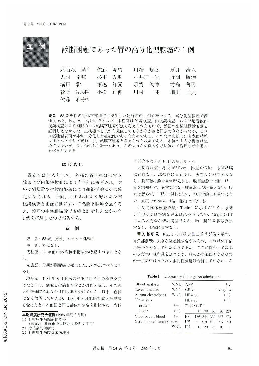
要旨 53歳男性の胃体下部前壁に発生した進行癌の1例を報告する.高分化型腺癌で深達度ssβ,ly2,V0,n1(+)であった.本症例はX線検査,内視鏡検査,および超音波内視鏡検査により肉眼的には粘膜下腫瘍が強く考えられたもので,頻回の生検組織診も癌を証明しえなかった.生検標本を後から見直してもなかなか癌と同定できなかったが,これは癌腫瘤表面が非常に分化した組織像であったためである.このため肉眼的にも表面粘膜はほとんど正常と変わらず,粘膜下腫瘍と考えられた次第である。本例のような胃癌は極めて少ないが,最近類似した報告もあり,このような症例も念頭に置いて胃癌診断を進めるべきと考える.
A 53-year-old man was referred to our hospital for further examination of the stomach. Physical and laboratory examinations (Table 1) revealed no abnormalities except for diabetes mellitus.
Radiological (Figs. 1 and 2), endoscopic (Fig. 3), and imaging (Fig. 4) examinations demonstrated a submucosal-tumor-like protruding lesion with some whitely coated depressions on the surface in the anterior wall of the lower gastric body. Biopsy sepcimens taken from the surface mucosa showed almost normal epithelium (Fig. 5) without any signs suggestive of malignancy.
The surgically obtained specimen (Fig. 6) also looked like a submucosal tumor, but the histological studies (Fig. 7) provided the evidences of carcinoma, i.e., tub1, ssβ, ly2, v0, n1(+).
Thus extremely well differentiation of the carcinoma in this patient presented the surface mucosa almost normal macroscopically and microscopically (in biopsy) as well. Although this type of carcinoma is very rare, we would like to emphasize the necessity to take it into consideration when making differential diagnosis of a gastric lesion.

Copyright © 1989, Igaku-Shoin Ltd. All rights reserved.


