Japanese
English
- 有料閲覧
PPV購入案内
1,320円 (1,200円+税10%) こちらは、341ページから347ページの文献です。購入には医書.jp本会員登録が必要です。
会員登録完了後に改めて上記「購入サイトへ」を選択してください。
または「購入サイトへ」選択後に「購入ログイン」、「新規会員登録」の順に進んでください。
会員登録について詳しくはをご確認ください。
- Abstract 文献概要
- 1ページ目 Look Inside
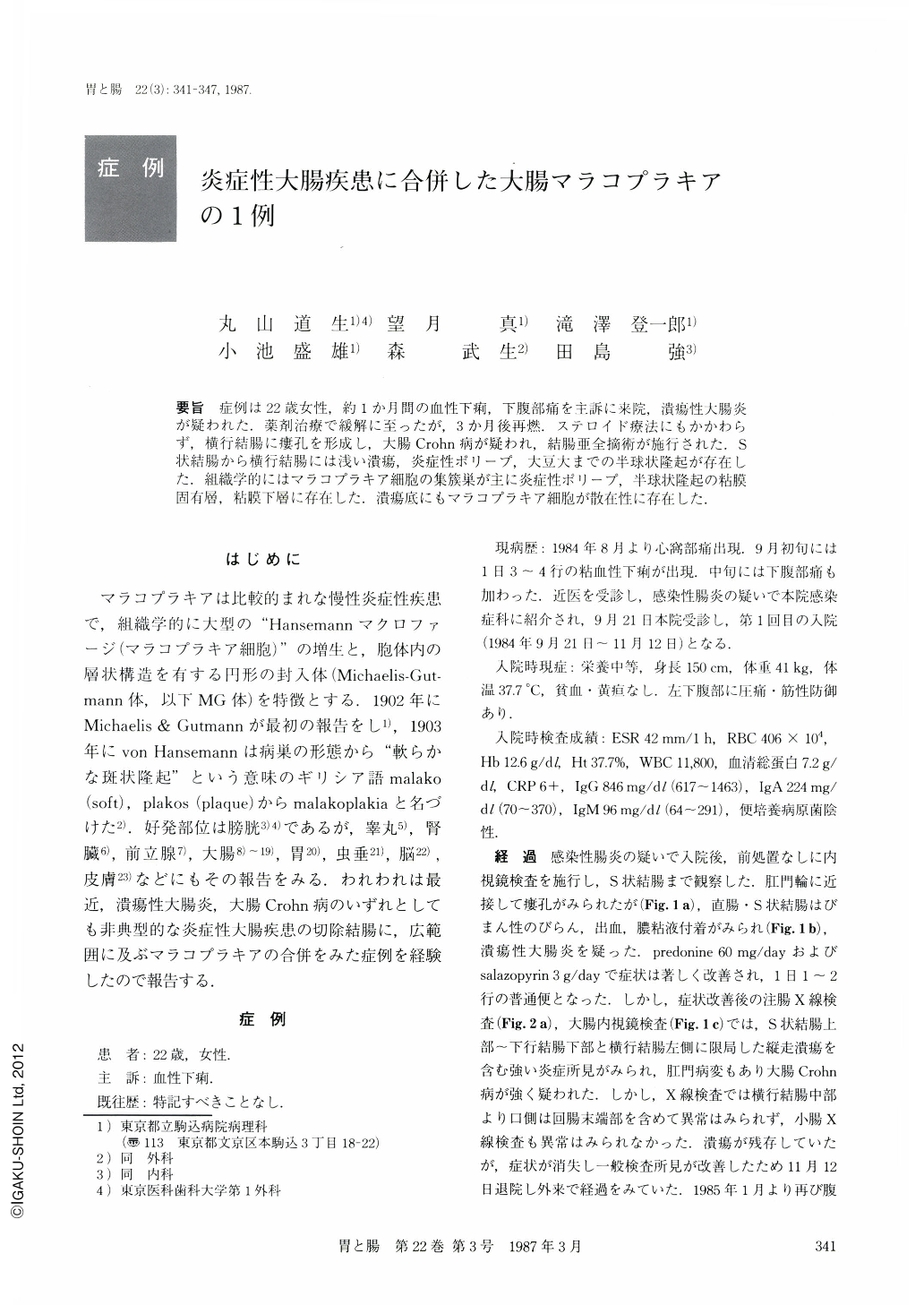
要旨 症例は22歳女性,約1か月間の血性下痢,下腹部痛を主訴に来院,潰瘍性大腸炎が疑われた.薬剤治療で緩解に至ったが,3か月後再燃.ステロイド療法にもかかわらず,横行結腸に瘻孔を形成し,大腸Crohn病が疑われ,結腸亜全摘術が施行された.S状結腸から横行結腸には浅い潰瘍,炎症性ポリープ,大豆大までの半球状隆起が存在した.組織学的にはマラコプラキア細胞の集蔟巣が主に炎症性ポリープ,半球状隆起の粘膜固有層,粘膜下層に存在した.潰瘍底にもマラコプラキア細胞が散在性に存在した.
Malakoplakia is a rare chronic inflammatory lesion which frequently affects the urinary bladder. We encountered extensive malakoplakia in the colon of a patient with inflammatory bowel disease which was not typical of ulcerative colitis or Crohn's disease.
The patient was a 22 year-old woman who was admitted to our hospital with complaints of bloody diarrhea and lower abdominal pain for the period of about one month. Clinically and endoscopically, ulcerative colitis was suspected. The symptoms were alleviated by the administration of salazopyrin and steroid. But three months later, she suffered from the same symptoms again. In spite of the steroid therapy for two months, fistular formation of the transverse colon was found. Biopy specimens before the operation revealed crypt abscesses, but neither granulomas nor malakoplakia cells. Subtotal colectomy was performed and postoperatively, the symptoms ceased.
Resected colon showed extensive shallow ulcerations, many tall pseudopolyps and up to soya-bean sized hemispherical elevated lesions. A fistula was observed at the transverse colon. On the cut surface, many yellowish foci were visible in the lamina propria and in the submucosal layer of the pseudopolyps and the hemispherical elevated lesions. Histologically, these foci were composed of accumulations of malakoplakia cells. Some of the cells contained intracytoplasmic spherical inclusion bodies,“Michaelis-Gutmann bodies”. And in addition, malakoplakia cells were scattered in the ulcer grounds. Many phagolysosomes amd laminated “Michaelis-Gutmann bodies” in the malakoplakia cells were observed by electronmicroscope. On the other hand, ulcerations were shallow and confined to the submucosal layer, except for a fistula of the transverse colon. There were crypt abscesses but no granulomas.
About 40 cases of gastrointestinal malakoplakia have been reported previously, and about 75% of these cases were colonic malakoplakia. Many cases were associated with malignant diseases. Only two cases of colonic malakoplakia associated with inflammatory bowel disease have been reported in the world medical literature.
Copyright © 1987, Igaku-Shoin Ltd. All rights reserved.