Japanese
English
- 有料閲覧
PPV購入案内
1,320円 (1,200円+税10%) こちらは、377ページから381ページの文献です。購入には医書.jp本会員登録が必要です。
会員登録完了後に改めて上記「購入サイトへ」を選択してください。
または「購入サイトへ」選択後に「購入ログイン」、「新規会員登録」の順に進んでください。
会員登録について詳しくはをご確認ください。
- Abstract 文献概要
- 1ページ目 Look Inside
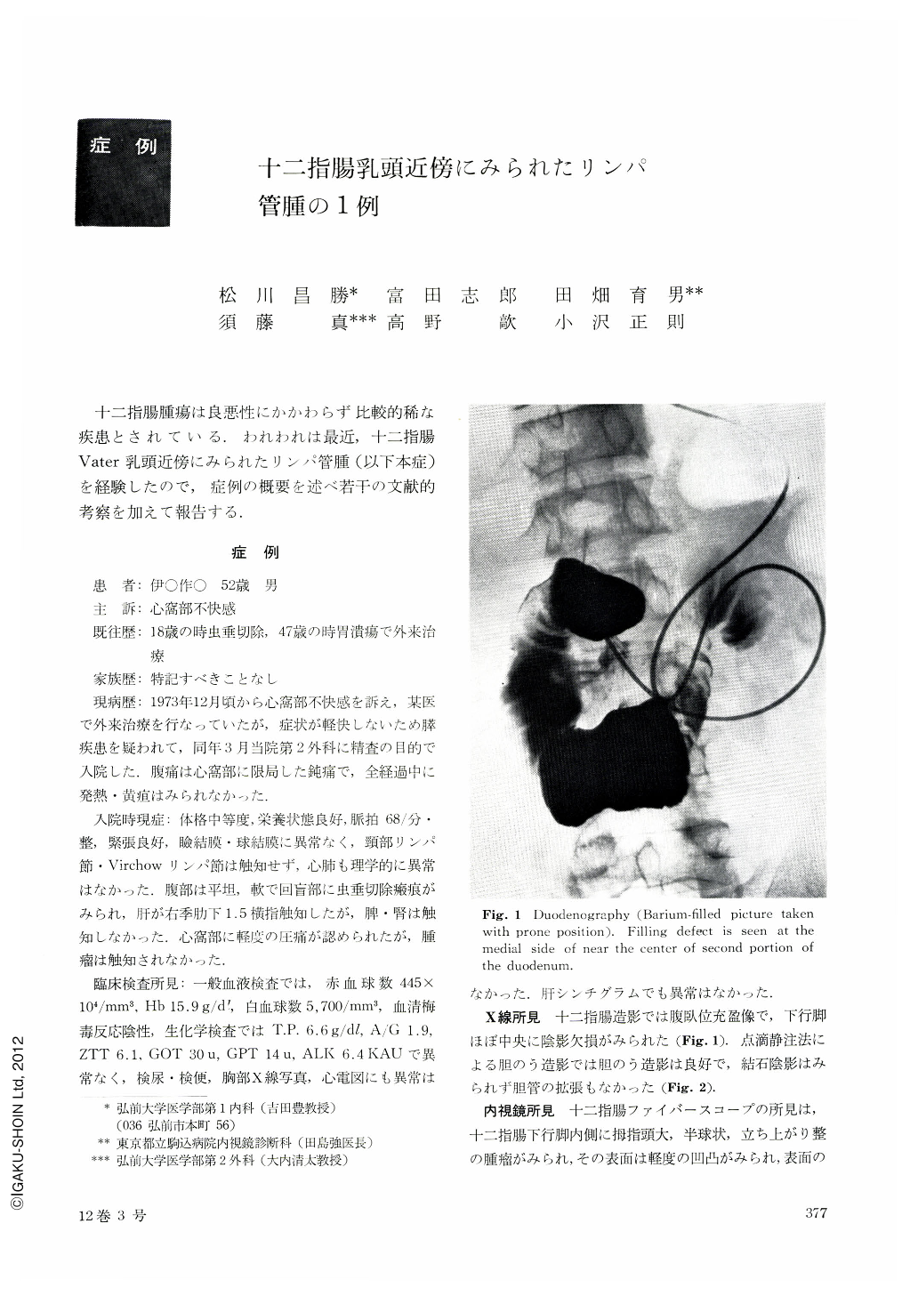
十二指腸腫瘍は良悪性にかかわらず比較的稀な疾患とされている.われわれは最近,十二指腸Vater乳頭近傍にみられたリンパ管腫(以下本症)を経験したので,症例の概要を述べ若干の文献的考察を加えて報告する.
A case with lymphangioma in the periampullary region is reported. The patient was a 52-year-old man whose chief complaint was epigastric pain. He had a past history of gastric ulcer at the age of 47. His family history was not contributory. He had begun to feel epigastric discomfort in December 1973 and had been treated at a private hospital without improvement of his discomfort. So he was admitted to the 2nd Department of Surgery of our hospital in March 1974 for thorough examination with a suspicion of pancreatic disorder. (His ache was a dull pain localized in epigastric area. No fever or icterus was noted throughout the disease.)
On physical examination at our hospital, an appendectomy scar was observed at right lower quadrant. The liver was palpable 1.5 finger breadths below the costal margin without any other abnormal physical findings.
With duodenography, a filling defect was seen at the medial side near the middle of second portion of the duodenum on the barium-filled picture taken with the prone position. With duodenofiberscopy, a tumor as big as a thumb-tip was found in the periampullary region at the medial side of second portion of the duodenum. Its surface was slightly jagged and a little more yellowish than its surrounding mucosa. With biopsy, small round-cell infiltration and slight fibrosis were observed in the submucosa but no heterokaryon was noticed.
Because of the difficulty to discriminate it from benign tumor of the papilla or from periampullary carcinoma, an abdominal operation was performed. (The second portion of the duodenum right above the papilla of Vater was incised, and a tumor as big as a thumb-tip was disclosed.) It was confirmed that the phyma was movable and its surface was compressible with palpation suggestive of a cystoid structure. When a curved needle with a thread was placed at the root of the tumor for the purpose of ligature, milky fluid came out, and the tumor collapsed, its cyst wall was excised and the operation was over after correction of defective mucosa.
Histological findings of the cyst wall revealed formation of numerous cavernous cavities lined with endothelium at the lower layer of mucosa. Final pathological diagnosis was made as lymphangioma cavernosum probably caused by the local stagnation of lymph.
Eventually the patient has been relieved from his discomfort after operation.
Copyright © 1977, Igaku-Shoin Ltd. All rights reserved.