Japanese
English
- 有料閲覧
PPV購入案内
1,320円 (1,200円+税10%) こちらは、463ページから467ページの文献です。購入には医書.jp本会員登録が必要です。
会員登録完了後に改めて上記「購入サイトへ」を選択してください。
または「購入サイトへ」選択後に「購入ログイン」、「新規会員登録」の順に進んでください。
会員登録について詳しくはをご確認ください。
- Abstract 文献概要
- 1ページ目 Look Inside
われわれは胃潰瘍の経過観察中に,約3ヵ月で大きな腫瘤を形成した胃平滑筋肉腫を経験したので報告する.
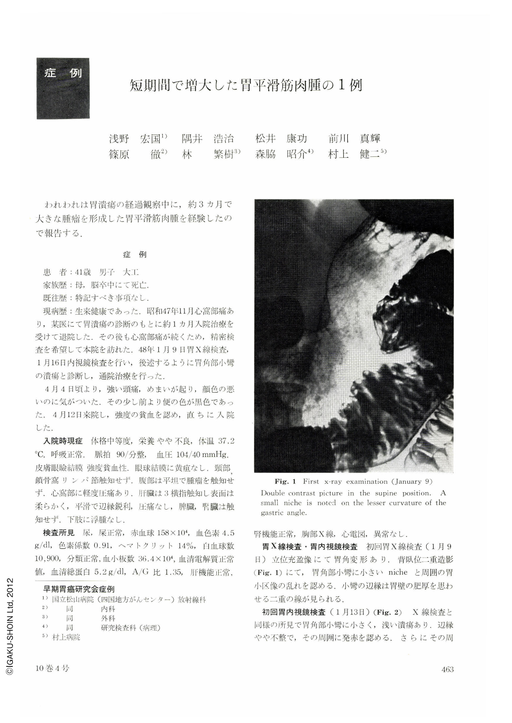
Complaining of epigastralgia for the last two momths, patient, 41-year-old, male, visited our out patient clinic. On Jan. 9, 1973 the upper G-I series and endoscopical examination revealed a small niche surrounded by minimal elevation of mucosa at the angulus of lesser curvature. He was treated for the niche for the following three months, when he began to complain of hematemesis and tarry stool off and on.
Emergently admitted on April 12, 1973, he was received the second upper G-I series, 97 days after the first examination and was detected a large tumor with nodular surface, sized 6.5×5.0 cm, at the angulus of lesser curvature. Biopsy revealed necrotic masses with no malignant cells.
The tumor was nodulated with bleeding and necrosis, sized 7.0×5.5×4.0 cm on the resected stomach. Two ulcerations was cleared, one was next to the tumor, and the other was crepyloric area. Histological examination revealed Leiomyosarcoma with infiltration into the subserosa. No metastasis was cleared (P0, H0, n0, S0, Ly0, V0).
The rapid growth of this tumor looks consisted of vascular proliferation, bleeding and edema.
Copyright © 1975, Igaku-Shoin Ltd. All rights reserved.