Japanese
English
- 有料閲覧
- Abstract 文献概要
- 1ページ目 Look Inside
Konjetzny(1938)1)は臨床的に胃癌と鑑別が困難で,組織学的にリンパ系の増殖を主とする症例を,慢性胃炎の1特殊型“Lymphatisch-hyperplastischer Gastritis”として報告した.その後,Smith & Helwig2)がReactive lymphoid hyperplasiaとして42例を,Faris & Saltzstein3)がLymphoid hyperplasiaとして21例を,また中村ら4)5)がReactive lymphoreticular hyperplasia(以下RLHと略す)として6例を総説し,以来散発的ながらかかる症例の報告がみられている.
最近,筆者らは胃体部小彎付近の限局性小病変で,レ線的・内視鏡的にⅡa+Ⅱc型早期胃癌を疑い,生検によりRLHを考え,胃切除により確認した1例を経験したので報告する.
Case: a 48-year-old woman visited the authors' clinic complaining of feeling of fullness in the stomach and hunger pain both lasting for two months. Previous history revealed no definite illness. Slight hypoaciditiy of the gastric juice was found at clinical examinations; otherwise blood and biochemical examinations were within normal limits.
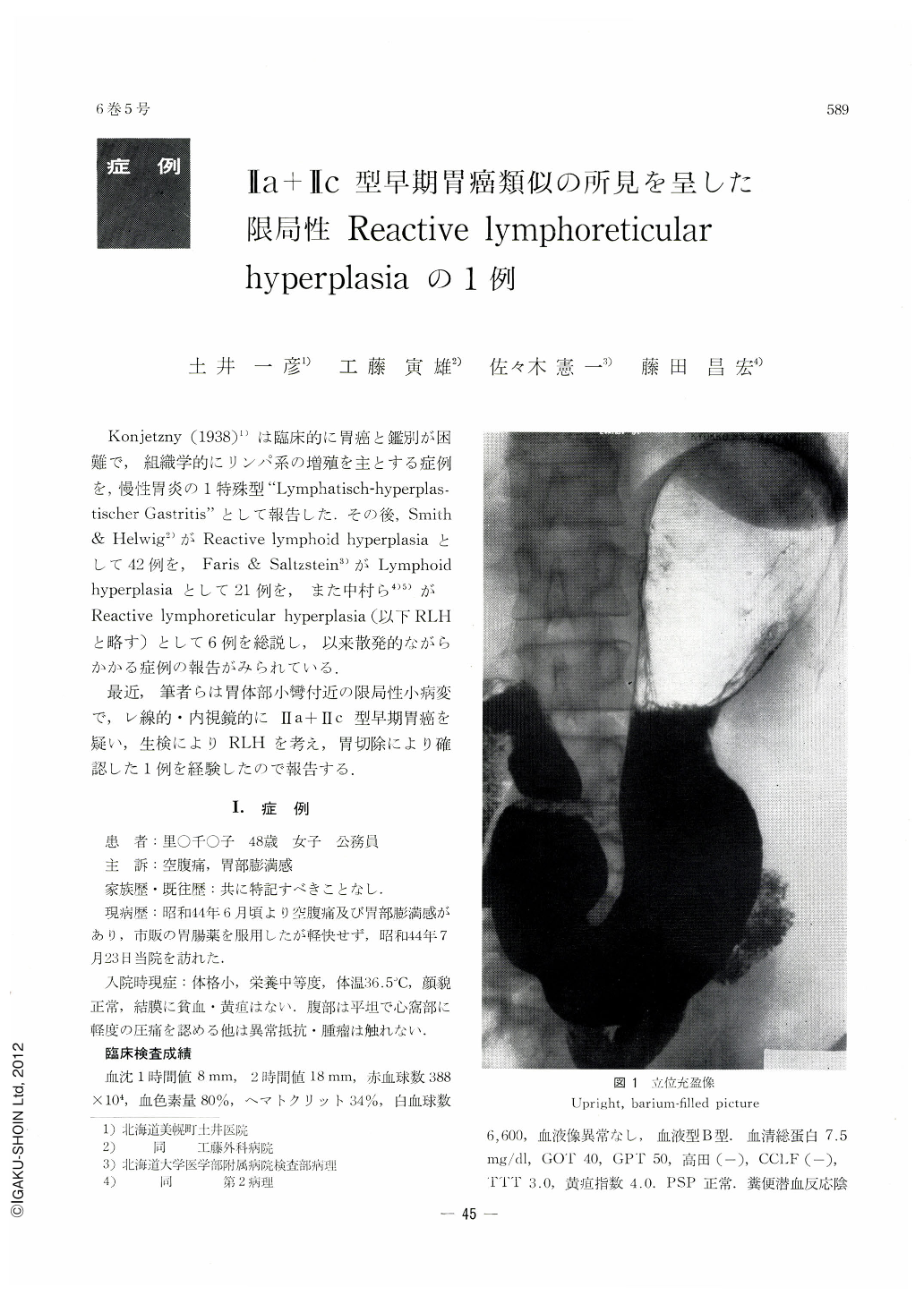
At x-ray examination of the stomach, a shadow looking like a Ⅱa lesion was visualized on the lesser curvature above the gastric angle in supine double contrast picture. The shadow was seen as a shallow niche by prone double contrast study. Endoscopically, a shallow depression of irregular shape with engorgement was revealed on the anterior wall near the lesser curvature of the upper corpus. Slight elevation was seen around the depression, but mucosal uprising was not so steep, and the tips of the mucosal folds, though swollen and ceased at the border of the depression, was relatively smooth.
Resected stomach revealed a shallow clepressed area of irregular shape, measuring 12×6 mm on the anterior wall side above the gastric angle. It was associated with ill-defined slight elevation.
Histologically, relatively sharply circumscribed, proliferated nests of lymphoreticular cells, extending from the mucosal to submucosal layers, were seen at a site corresponding to the depression and its encircling elevation. In addition to infiltration by plasma cells and neutrophile cells, formation of lymph follicles having central reaction was manifest. As no malignant finding was seen, this case was diagnosed as reactive lymphoreticular hyperplasia of the stomach.
The muscularis mucosae at the site corresponding to the lesion was irregular and in looser arrangement. The muscle fibers were partly discontinued, but overall changes were slight.
RLH is usually found after considerably long clinical course in association with ulcer lesion. The case described here belongs to RLH detected in its fairly early stage.

Copyright © 1971, Igaku-Shoin Ltd. All rights reserved.


