Japanese
English
- 有料閲覧
PPV購入案内
1,320円 (1,200円+税10%) こちらは、77ページから80ページの文献です。購入には医書.jp本会員登録が必要です。
会員登録完了後に改めて上記「購入サイトへ」を選択してください。
または「購入サイトへ」選択後に「購入ログイン」、「新規会員登録」の順に進んでください。
会員登録について詳しくはをご確認ください。
- Abstract 文献概要
- 1ページ目 Look Inside
Ⅰ.はじめに
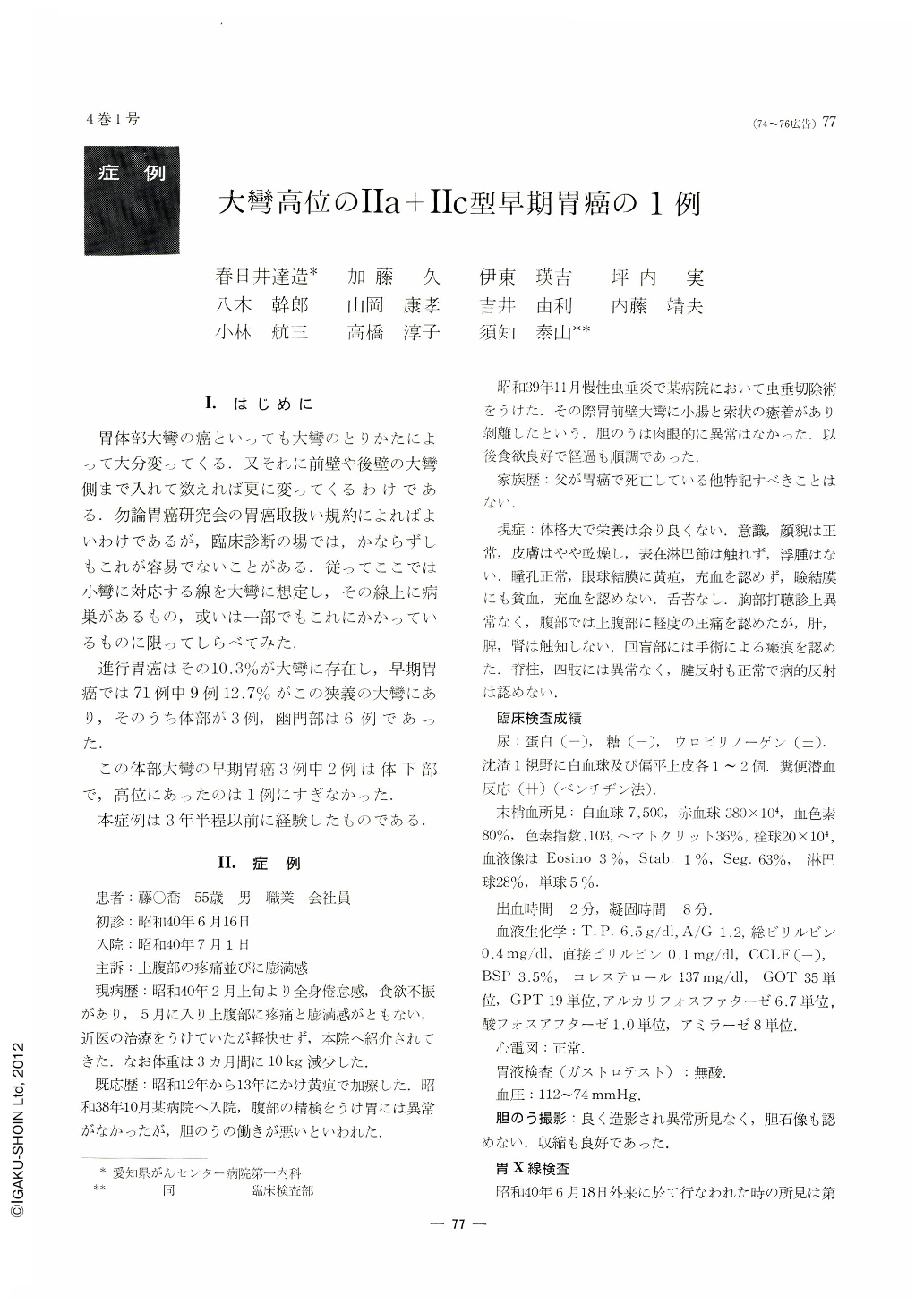
胃体部大彎の癌といっても大彎のとりかたによって大分変ってくる.又それに前壁や後壁の大彎側まで入れて数えれば更に変ってくるわけである.勿論胃癌研究会の胃癌取扱い規約によればよいわけであるが,臨床診断の場では,かならずしもこれが容易でないことがある.従ってここでは小彎に対応する線を大彎に想定し,その線上に病巣があるもの,或いは一部でもこれにかかっているものに限ってしらべてみた.
進行胃癌はその10.3%が大彎に存在し,早期胃癌では71例中9例12.7%がこの狭義の大彎にあり,そのうち体部が3例,幽門部は6例であった.
この体部大彎の早期胃癌3例中2例は体下部で,高位にあったのは1例にすぎなかった.本症例は3年半程以前に経験したものである.
A 55-year-old man was admitted to the hospital because of complaints of epigastralgia and full sensation of the stomach for about five months. Hematologic examination showed RBC of 3.80 million, Hgb 80% (Sahli) and WBC 7,500. Occult blood in feces was moderately positive. Gastric analysis revealed achylia. X-ray examination of the stomach revealed a niche shaped “Schattenplus im Schattenminus” on the greater curvature in the upper gastric body.
Endoscopy showed a slightly protruded lesion with an irregular concavity in its center like the Ⅱa+Ⅱc type of early gastric cancer on the greater curvature of the upper gastric body, as well as three ulcers, one of which was small and located on the angulus, and the others moderate sized, one located on the anterior wall and another on the posterior wall near the greater curvature in the upper gastric body respectively.
Gastric cytology under direct vision by the fibergastroscope was positive, however, gastric biopsy under direct vision failed to show a cancer in successful biopsy. Gastrectomy by the method of Billroth I was carried out. The resected specimen had an irregularly shaped, protruded lesion with a slight depression on its surface, measuring 15×20 mm in diameter just on the greater curvature of the upper gastric body, and two ulcers in the upper gastric body. Histological examination of the former revealed a well differentiated adenocarcinoma, involving only the mucosal and submucosal layers. Pathological classification was adenocarcinoma tubulare, CATI, SAT1, Inf. α, sm.
Copyright © 1969, Igaku-Shoin Ltd. All rights reserved.