Japanese
English
- 有料閲覧
- Abstract 文献概要
- 1ページ目 Look Inside
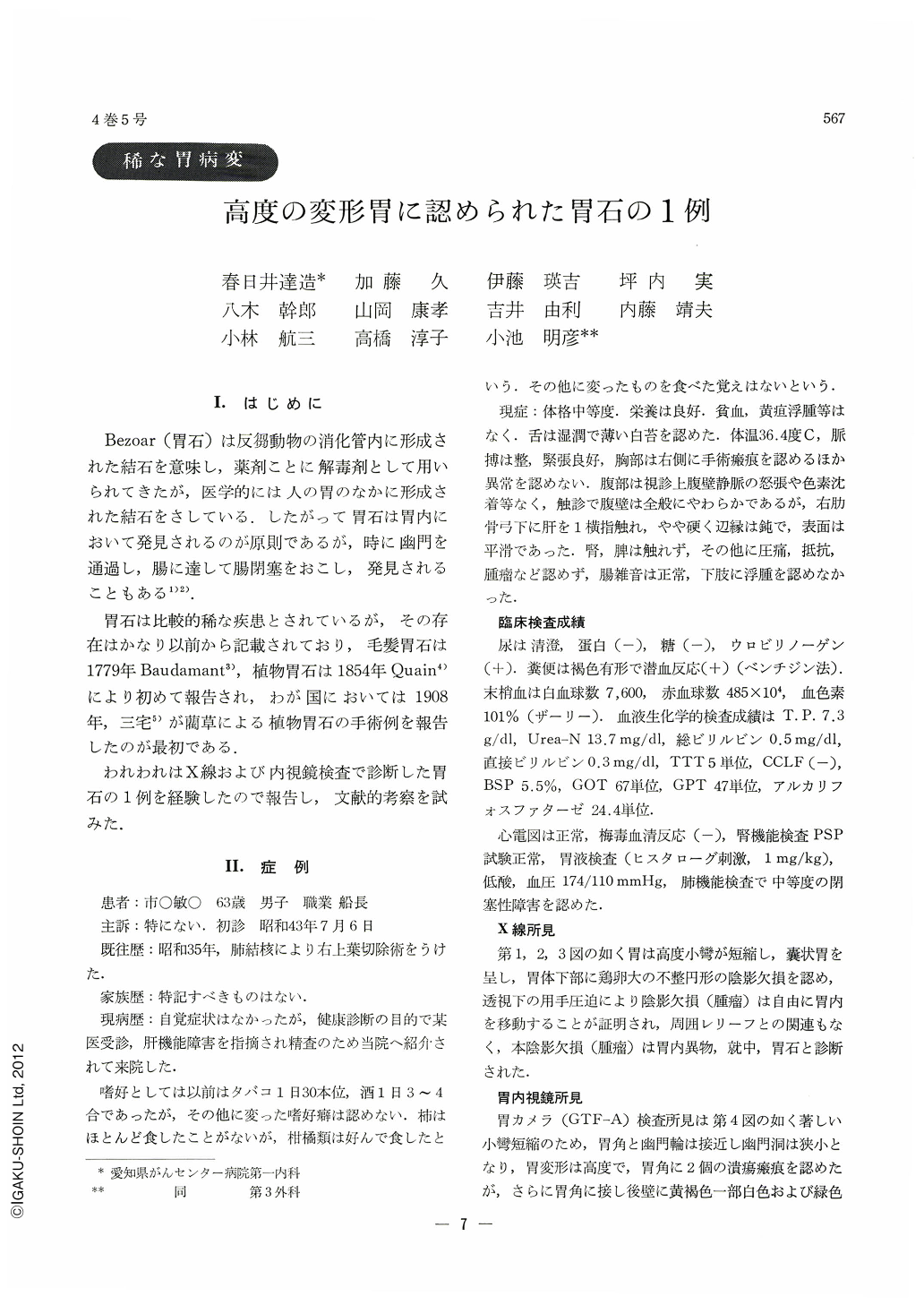
Ⅰ.はじめに
Bezoar(胃石)は反胃動物の消化管内に形成された結石を意味し,薬剤ことに解毒剤として用いられてきたが,医学的には人の胃のなかに形成された結石をさしている.したがって胃石は胃内において発見されるのが原則であるが,時に幽門を通過し,腸に達して腸閉塞をおこし,発見されることもある1)2).
胃石は比較的稀な疾患とされているが,その存在はかなり以前から記載されており,毛髪胃石は1779年Baudamant3),植物胃石は1854年Quain4)により初めて報告され,わが国においては1908年,三宅5)が藺草による植物胃石の手術例を報告したのが最初である.
われわれはX線および内視鏡検査で診断した胃石の1例を経験したので報告し,文献的考察を試みた.
T. I. A 63-year-old man who had impairment of the liver function but no specific complaint was admitted to hospital for further follow-up.
X-ray examination of the stomach revealed marked shortening of the lesser curvature and marked deformity of the stomach, and an irregularly shaped, egg-size round filling defect which could be moved within the stomach by manipulation. No mucosal rugae related to the filling defect could be demonstrated.
Gastrocamera (GTF-A) pictures revealed an irregularly shaped large globular mass with yellowbrown, partially white and green color, and with nodularly rough surface on the posterior wall just near the angulus, and marked shortening of the lesser curvature, marked deformity of the stomach, and two ulcer-scars on the angulus.
The mass moved from the lower gastric body to the fornix and half sunk into the mucous lake when position of the patient was changed. This case was diagnosed to have a bezoar and ulcer-scars endoscopically.
The mass was confirmed as a bezoar by the microscopical examination of some small fragments taken by forceps incorporated in the fibergastroscopy for biopsy. Colonies of candida albicans as proved by culture were also demonstrated on them.
The bezoar with a size of 5 × 4 × 3 cm, weighing 22.5 g was removed by gastrotomy. The bezoar, a potato-like mass, showed violet, red-brown in color. Its surface was nodularly rough with partially grey-white adhered stuff like mold, and cork-like elastic softness. Hair ball, skin, pulp or seeds of the vegetable were not demonstrated on the section of the bezoar, and microscopic examination demonstrated the mass consisting of vegetable fibers with adhered mucous substances and marked propagation of candida albicans on or in the bezoar.
The bezoar was presumed as a kind of iniobezoar.
Main causes of the bezoar in this case were probably much eating of cisrus-fruits, delayed emptying resulting from the deformity of the stomach due to multiple ulcer-scars and the shortening of the lesser curvature, and the congestion in the stomach due to liver cirrhosis.
Copyright © 1969, Igaku-Shoin Ltd. All rights reserved.