Japanese
English
- 有料閲覧
PPV購入案内
1,320円 (1,200円+税10%) こちらは、935ページから947ページの文献です。購入には医書.jp本会員登録が必要です。
会員登録完了後に改めて上記「購入サイトへ」を選択してください。
または「購入サイトへ」選択後に「購入ログイン」、「新規会員登録」の順に進んでください。
会員登録について詳しくはをご確認ください。
- Abstract 文献概要
- 1ページ目 Look Inside
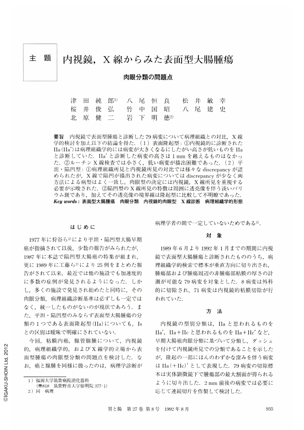
要旨 内視鏡で表面型腫瘍と診断した79病変について病理組織との対比,X線学的検討を加え以下の結論を得た.(1)表面隆起型:①内視鏡的に診断されたⅡa(Ⅱa’)は病理組織学的には病変が大きくなるにしたがい高さが低いものをⅡaと診断していた.Ⅱa’と診断した病変の高さは1mmを越えるものはなかった.②ルーチンX線検査では小さく,低い病変が描出困難であった.(2)平坦・陥凹型:①病理組織所見と内視鏡所見の対比では様々なdiscrepancyが認められたが,X線で陥凹が描出された病変についてはdiscrepancyが少なく両方法による病型はよく一致し,肉眼型の決定には内視鏡,X線所見を重視する必要が示唆された.②陥凹型のX線所見の特徴は周囲に透亮像を伴う淡いバリウム斑であり,加えてその透亮像の境界線は隆起型に比較して不明瞭であった.
Endoscopic, radiographic and pathomorphologic comparison was performed on 79 superficial colonic neoplastic lesions originally diagnosed by colanoscopy. Results are as follows:
1) Superficial elevated lesions; i) The endoscopic diagnosis of Ⅱa was frequently made as the size of the lesion was bigger, and the height was lower on the resected specimen. However, there was no lesion higher than 1 mm. ii) It was difficult to demonstrate the small lesions in size and height.
2) Flat and depressed lesions; i) There were various discrepancies between the endoscopic and pathomorphologic findings. However, the radiologically demonstrated lesions coincided with the endoscopic and pathomorphologic findings, therefore both endoscopic and radiologic results was important in determining morphology. ii) The radiologic characteristics of the depressed type was a barium fleck accompanied by surrounding translucency. The border of the traslucent area of depressed type was less clear than that of the elevated type.
Copyright © 1992, Igaku-Shoin Ltd. All rights reserved.