Japanese
English
- 有料閲覧
PPV購入案内
1,320円 (1,200円+税10%) こちらは、879ページから888ページの文献です。購入には医書.jp本会員登録が必要です。
会員登録完了後に改めて上記「購入サイトへ」を選択してください。
または「購入サイトへ」選択後に「購入ログイン」、「新規会員登録」の順に進んでください。
会員登録について詳しくはをご確認ください。
- Abstract 文献概要
- 1ページ目 Look Inside
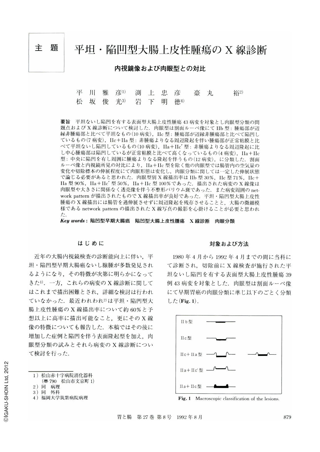
要旨 平坦ないし陥凹を有する表面型大腸上皮性腫瘍43病変を対象とし肉眼型分類の問題点およびX線診断について検討した.肉眼型は割面ルーペ像にてⅡb型:腫瘍部が辺縁非腫瘍部と比べて平坦なもの(10病変),Ⅱc型;腫瘍部が辺縁非腫瘍部と比べて陥凹しているもの(7病変),Ⅱc+Ⅱa型:非腫瘍よりなる周辺隆起を伴い腫瘍部が正常粘膜と比べて平坦ないし陥凹しているもの(10病変),Ⅱa+Ⅱc’型:非腫瘍よりなる周辺隆起に比し中心腫瘍部は陥凹しているが正常粘膜と比べて高くなっているもの(4病変),Ⅱa+Ⅱc型:中央に陥凹を有し周囲に腫瘍よりなる隆起を伴うもの(12病変),に分類した.割面ルーペ像と内視鏡所見の対比により,Ⅱa+Ⅱc型を除く他の肉眼型では腸管内の空気量の変化や切除標本の伸展程度にて肉眼形態は変化し,肉眼分類に関しては一定した伸展状態で論じる必要があると思われた.肉眼型別X線描出率はⅡb型30%,Ⅱc型71%,Ⅱc+Ⅱa型90%,Ⅱa+Ⅱc’型50%,Ⅱa+Ⅱc型100%であった.描出された病変のX線像は肉眼型や大きさに関係なく透亮像を伴う不整形バリウム斑であった.また病変周囲のnetwork pattemが描出されたものでX線描出率が良好であった.平坦・陥凹型大腸上皮性腫瘍のX線描出には腸管を過伸展させずに周辺隆起を残存させることと,大腸の微細模様であるnetwork patternの描出されたX線写真の撮影を心掛けることが必要と思われた.
We evaluated the macroscopic classification and radiologic diagnosis of 43 flat or depressed superficial epithelial tumors of the large intestine. Macroscopically they were classified as follows; 10 superficial flat (Ⅱb), 7 superficial depressed without marginal elevation (Ⅱc), 10 superficial depressed with marginal elevation consisting of non-neopiastic tissue (Ⅱc+Ⅱa), 4 slight elevation with marginal elevation consisting of non-neoplastic tissue (Ⅱa+Ⅱc'), and 12 depressed with marginal elevation consisting of tumor (Ⅱa+Ⅱc). The macroscopic classification should be discussed under the same grade of extension of the bowel wall because the macroscopic appearance of the superficial epithelial tumors, except for Ⅱa+Ⅱc type, changed with pneumatic distention. The radiological detectability differed from macroscopic shape in 30% (Ⅱb), 71% (ⅡC), 90% (Ⅱc+Ⅱa), 50% (Ⅱa+Ⅱc'), and 100% (Ⅱa+Ⅱc), respectively. The radiologic finding of these detectable lesions was an irregularly shaped barium fleck with surrounding radiolucency, irrespective of macroscopic shape and size of the tumor. It was necessary to avoid pneumatic overextention in order to show marginal elevation and to prepare the x-ray equipment so as to demonstrate the surrounding network pattern of normal mucosa. In this way, it was possible to detect flat or depressed epithelial tumor of the large intestine.
Copyright © 1992, Igaku-Shoin Ltd. All rights reserved.