Japanese
English
- 有料閲覧
PPV購入案内
1,320円 (1,200円+税10%) こちらは、1097ページから1105ページの文献です。購入には医書.jp本会員登録が必要です。
会員登録完了後に改めて上記「購入サイトへ」を選択してください。
または「購入サイトへ」選択後に「購入ログイン」、「新規会員登録」の順に進んでください。
会員登録について詳しくはをご確認ください。
- Abstract 文献概要
- 1ページ目 Look Inside
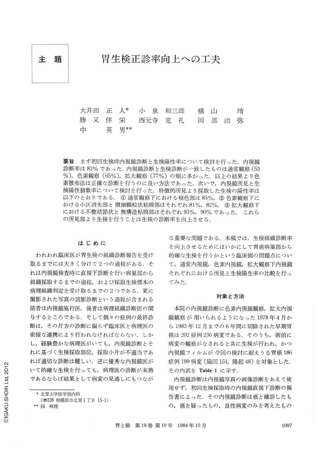
要旨 まず初回生検時内視鏡診断と生検陽性率について検討を行った.内視鏡診断率は83%であった.内視鏡診断と生検診断が一致したものは通常観察(53%),色素観察(65%),拡大観察(77%)の順に多かった.以上の結果より色素散布法は正確な診断を行うのに良い方法であった.次いで,内視鏡所見と生検陽性個数率について検討を行った.特徴的所見より採取した生検の陽性率は以下のとおりである.①通常観察下における褪色部は85%,②色素観察下における小区消失部と微細顆粒状粘膜部はそれぞれ81%,82%,③拡大観察下における不整結節状と無構造粘膜部はそれぞれ93%,90%であった.これらの所見部より生検を行うことは生検の診断率を向上させる.
At first, we studied early gastric cancers on initial endoscopic diagnosis with biopsy and their positive biopsy ratio.
Endoscopic diagnostic ratio was 83%. Diagnostic ratio coincided with endoscopic and biopsy findings was increased using conventional (53%), dye scattering (65%) and magnifying endoscopic method (77%) in turn. As a result, dye scattering was the most reliable method for accurate diagnosis.
Next, we investigated the relationship between endoscopic findings and the ratio of positive number of biopsy specimens. Positive ratio of biopsy specimens obtained from the characteristic findings was as follows: 1) Discolored area under the conventional endoscopy was 85%. 2) Disappeared area and fine granular area under the dye scattering method were 81% and 82%, respectively. 3) Irregular nodule-like mucosa and flattened mucosa under the magnifying endoscopy were 93% and 90%, respectively. Taking biopsy specimens from these areas is recommended for the increase of the diagnostic ratio of biopsy.
Copyright © 1984, Igaku-Shoin Ltd. All rights reserved.