Japanese
English
- 有料閲覧
PPV購入案内
1,320円 (1,200円+税10%) こちらは、203ページから206ページの文献です。購入には医書.jp本会員登録が必要です。
会員登録完了後に改めて上記「購入サイトへ」を選択してください。
または「購入サイトへ」選択後に「購入ログイン」、「新規会員登録」の順に進んでください。
会員登録について詳しくはをご確認ください。
- Abstract 文献概要
- 1ページ目 Look Inside
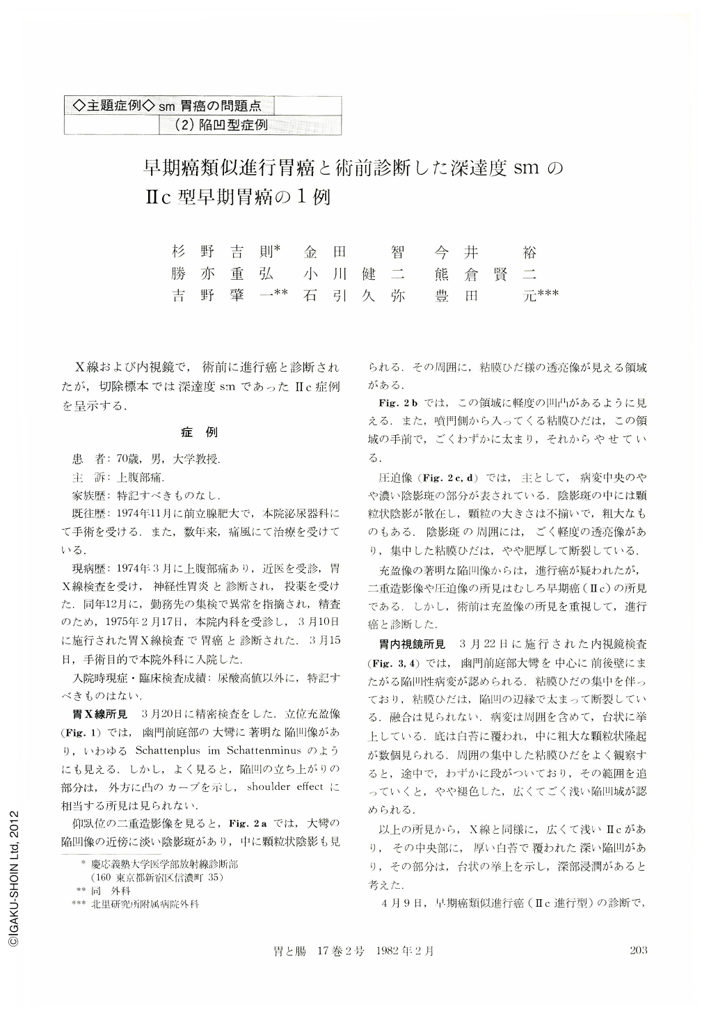
X線および内視鏡で,術前に進行癌と診断されたが,切除標本では深達度smであったⅡc症例を呈示する.
症例
患 者:70歳,男,大学教授.
主 訴:上腹部痛.
家族歴:特記すべきものなし.
既往歴:1974年11月に前立腺肥大で,本院泌尿器科にて手術を受ける.また,数年来,痛風にて治療を受けている.
The patient, a 70-year-old man, complained of epigastralgia since March 1974. Routine upper GI series performed on March 10, 1975, revealed an abnormality suggesting gastric cancer. Preoperatively he was diagnosed to have advanced cancer simulating type Ⅱc by means of minute x-ray examination and endoscopic study. Findings were marked deformity of the antrum and double-contoured depressed lesion, and this depressed area was noticed in plateau-like elevation. Partial gastrectomy was carried out on April 9, 1975. Histological examination was proved to be undifferentiated adenocarcinoma. The size of the lesion was 6.5×3.5 cm and the lesion had double contour. A linear scar (Ul-Ⅱ) was identified within the inner, deeper depression. Cancerous involvement was limited in the submucosal layer.
Retrospectively, the marked deformity of the greater curvature and the plateau-like elevation were considered to be caused by the linear scar and submucosal fibrosis. If the linear scar had been recognized, preoperatively we might have been able to diagnose this patient as early gastric cancer.
Copyright © 1982, Igaku-Shoin Ltd. All rights reserved.